
Bioprinting Quality Intelligence: Lab-Printed Human Tissue Will Function Properly

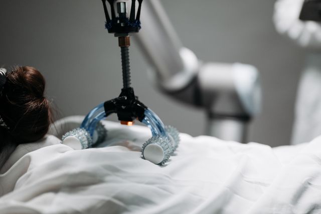
Advances in biotechnology are changing how scientists approach the development of human tissues for medical research, drug testing and regenerative medicine. One of the most promising developments is three-dimensional bioprinting, a technique that uses specialised printers to arrange living cells and biomaterials into structures that resemble biological tissues. Unlike conventional 3D printing, which generally works with plastics, metals or other non-living materials, bioprinting involves carefully positioning living cells within supportive materials known as bioinks.
The technology has created new possibilities for producing tissue models that can help researchers study diseases, examine how cells respond to medicines and investigate potential approaches to repairing damaged organs. However, printing a tissue structure that looks anatomically convincing does not necessarily mean that it will function like natural human tissue. Cells must remain alive, communicate with neighbouring cells, develop appropriate organisation and perform the biological activities expected of the target tissue.
This creates a major quality-control challenge. Researchers need to determine not only whether a printed structure has the correct shape but also whether its cells are healthy, its internal organisation is appropriate and its biological functions remain stable. Traditional quality assessment may involve microscopy, laboratory assays and repeated experiments, which can be time-consuming and may not capture every change occurring within a developing tissue.
Bioprinting Quality Intelligence explores how artificial intelligence could help predict the quality and functionality of lab-printed human tissues. By analysing imaging data, printing conditions, cellular behaviour and biological measurements, AI may help researchers identify potential problems earlier and improve the consistency of bioprinted structures. The central question is whether machine learning can move tissue engineering beyond visual inspection towards more comprehensive predictions of biological performance.
Understanding 3D Bioprinting and Human Tissue Engineering
Three-dimensional bioprinting is an additive manufacturing process in which living cells and biomaterials are deposited in controlled patterns to create biological structures. The process generally involves designing a digital model, preparing a suitable bioink and using a printing system to position the material layer by layer.
Bioinks are particularly important because they provide an environment in which cells can survive and develop. They may contain natural or synthetic polymers, hydrogels, growth-supporting components and living cells. The material must have suitable physical properties for printing while also supporting cellular viability and biological activity.
Different bioprinting techniques use different methods to deposit these materials. Extrusion-based systems push bioink through a nozzle, while inkjet-based systems deposit small droplets. Light-assisted and laser-based approaches use other mechanisms to position or solidify biological materials. Each method has advantages and limitations related to printing resolution, cell compatibility, material viscosity and production speed.
The intended tissue determines many of the design requirements. A skin model needs an appropriate layered structure, while cartilage requires mechanical properties that support its role in joints. Vascular tissue presents additional challenges because it must support the movement of blood or suitable fluid through internal channels.
The objective is not simply to reproduce the external appearance of a tissue. A successful bioprinted model must demonstrate biological characteristics relevant to its intended application. This distinction makes quality intelligence essential to the future development of bioprinting.
Why Structural Accuracy Does Not Guarantee Biological Function
One of the most important challenges in bioprinting is the difference between structural quality and functional quality. A printed tissue may have the correct dimensions, shape and visible organisation but still contain unhealthy or poorly functioning cells.
For example, a bioprinted liver-like tissue model may resemble a small section of liver under a microscope. However, its value for research depends on whether the cells maintain relevant metabolic activities, produce appropriate proteins and respond to chemical substances in ways that resemble the biological processes being studied.
Similarly, a printed cartilage model may have a suitable external shape but insufficient mechanical strength or an inadequate distribution of cells. A vascular structure may contain channels that look correct but fail to support stable fluid movement because the internal surfaces are not properly developed.
These differences make conventional visual inspection insufficient. Researchers need to examine several dimensions of quality, including cell survival, tissue organisation, mechanical stability, metabolic activity and the ability to perform specific biological functions.
AI could help connect these measurements. Instead of evaluating each quality indicator separately, machine learning models may identify relationships between printing conditions, structural characteristics and later biological performance. This could allow researchers to estimate whether a newly printed tissue is likely to meet its intended requirements.
How AI Can Predict Bioprinting Quality
Bioprinting systems generate large quantities of information during production and subsequent tissue development. This information may include printing speed, nozzle pressure, temperature, material composition, cell density, image measurements and laboratory test results.
Artificial intelligence can analyse these data sources to identify patterns associated with successful or unsuccessful tissue development. Machine learning models can be trained using previously printed samples for which researchers have recorded both production conditions and biological outcomes.
For instance, a model may learn that certain combinations of printing pressure, bioink viscosity and nozzle movement are associated with reduced cell viability. It could then assess new printing configurations and estimate whether they carry a higher risk of damaging cells.
Image-analysis models may examine the distribution of cells, the shape of printed layers and the consistency of internal structures. Other models could analyse laboratory measurements related to metabolism, protein production or tissue-specific activity.
Predictive models may also combine information collected at different stages. Early measurements taken immediately after printing could be compared with later observations to estimate whether a tissue is likely to remain stable during maturation.
However, AI predictions are only as dependable as the data and validation methods behind them. A model trained on one bioink, printer or cell type may not work reliably with another. Its predictions must therefore be tested across different materials, manufacturing conditions and biological applications.
Cell Viability Intelligence: Predicting Whether Printed Cells Will Survive
Cell viability is one of the most fundamental indicators of bioprinting quality. During printing, cells may experience mechanical stress, pressure changes, temperature variations and exposure to materials that affect their survival. If too many cells are damaged during the process, the resulting tissue may fail to develop the intended biological properties.
AI could help predict cell viability by analysing printing conditions alongside imaging and experimental measurements. For example, models could examine how nozzle diameter, extrusion pressure and printing speed relate to cell survival in a particular bioink.
Microscopy images may also provide useful information. Computer vision systems can identify cell shapes, estimate cell density and detect visible patterns associated with damaged or poorly distributed cells. When combined with validated viability assays, these measurements may help build more detailed quality assessments.
The timing of measurement is important. A cell may remain alive immediately after printing but become unhealthy during the following days because of inadequate nutrient supply, unsuitable material conditions or other environmental stresses. Therefore, quality prediction should consider both immediate survival and longer-term cellular behaviour.
AI may help identify early warning patterns, but it cannot establish cell viability from appearance alone in every situation. Biological assays remain necessary to verify whether the model’s predictions correspond to actual cell survival and function.
Predicting Tissue Architecture and Cellular Organisation
The internal organisation of cells plays a major role in determining how a tissue functions. Natural tissues contain specialised arrangements of cells, extracellular matrix and supporting structures. These arrangements influence communication between cells, nutrient transport, mechanical behaviour and biological activity.
Bioprinted tissues must reproduce relevant aspects of this organisation. If cells are distributed unevenly or layers do not develop as expected, the tissue may show reduced performance even when its overall shape appears correct.
AI-powered image analysis could help assess cellular organisation at different scales. Computer vision models may examine microscopy images to estimate cell distribution, identify gaps and detect variations in tissue density. Three-dimensional imaging data could provide additional information about internal structures that are difficult to assess through surface observations.
For example, a model designed for cartilage engineering could examine whether cells are distributed consistently throughout the printed structure. A system developed for skin models could assess whether the expected layers have developed in an appropriate arrangement.
These measurements could be compared with established reference structures and functional outcomes. AI may then estimate whether the tissue’s organisation is consistent with the requirements of its intended application.
However, biological tissues are not always perfectly uniform. Natural variation may be normal, and excessive emphasis on visual consistency could lead to the rejection of useful tissue models. Quality systems must therefore distinguish between harmless variation and structural problems that affect function.
Monitoring Vascularisation and Nutrient Distribution
One of the most difficult challenges in producing larger bioprinted tissues is ensuring that cells receive sufficient oxygen and nutrients. In natural tissues, blood vessels support the transport of oxygen, nutrients and waste products. Without an adequate transport system, cells located deep inside a printed structure may become damaged or die.
Researchers are investigating different methods of incorporating vascular-like channels into bioprinted structures. These channels may eventually be developed into more complex vascular networks, although producing stable and functional networks remains challenging.
AI could help analyse images and experimental measurements associated with these structures. It may identify whether channels are correctly formed, estimate their dimensions and detect areas where the network appears incomplete. Computational models could also examine how channel geometry and material properties influence fluid movement.
In combination with experimental measurements, AI may help predict whether nutrients and oxygen can reach the relevant regions of a tissue model. This is especially important for larger structures, where transport limitations can reduce cell survival and biological performance.
However, predicting fluid movement through a printed channel is not equivalent to demonstrating a functional blood vessel. A biological vascular system requires additional characteristics, including appropriate cell lining, stability and compatibility with surrounding tissue. AI can support analysis, but experimental validation remains essential.
Bioink Intelligence: Finding the Right Material for Each Tissue
Bioink selection strongly influences the success of bioprinting. Materials must be printable, maintain their structure after deposition and provide an environment that supports cell survival and development. These requirements can sometimes conflict.
A bioink that is highly suitable for printing may be too stiff or biologically unsuitable for a particular cell type. A softer material may support cellular development but fail to maintain the required shape. Researchers must balance viscosity, mechanical properties, degradation behaviour and biological compatibility.
AI could help identify relationships between bioink composition and tissue outcomes. By analysing experimental datasets, models may estimate how different combinations of polymers, crosslinking conditions and additives influence printing performance and cellular behaviour.
Researchers could also use predictive systems to narrow down material combinations before conducting extensive laboratory experiments. This may reduce the time required to explore possible formulations and help identify promising candidates for further testing.
A more advanced approach could connect material predictions with tissue-specific requirements. A bioink intended for cartilage may need different mechanical characteristics from one designed for soft neural tissue. AI systems could compare the requirements of each application with the properties of candidate materials.
Nevertheless, material recommendations must be verified experimentally. Biological responses can be difficult to predict because they depend on interactions among cells, materials and environmental conditions.
Functional Testing: Can AI Predict What a Printed Tissue Will Actually Do?
The most important question in bioprinting quality intelligence is whether a printed tissue performs its intended biological function. This is more difficult to measure than physical dimensions or visible structure because different tissues carry out different activities.
For example, liver tissue models may be assessed through metabolic activity and the production of specific proteins. Cardiac tissue models may be evaluated for coordinated contraction and electrical behaviour. Cartilage models may be examined for mechanical properties and the production of relevant extracellular matrix components.
AI could analyse these functional measurements and identify combinations associated with successful tissue performance. Instead of relying on one isolated test, a model might evaluate several indicators together and estimate whether the tissue meets predefined functional criteria.
Longitudinal analysis could be particularly useful. A tissue model may show acceptable activity shortly after printing but gradually lose function during maturation. Repeated measurements could help AI identify trends that indicate whether the structure is developing as expected.
Such predictions may help researchers prioritise samples for further testing and identify production conditions associated with better outcomes. However, AI should not replace functional assays. It must first demonstrate that its predictions reliably correspond to experimentally measured biological performance.
Applications in Drug Testing and Disease Research
Bioprinted tissue models are increasingly important in research because they can provide controlled environments for studying biological processes. Researchers may use them to investigate how cells respond to drugs, environmental conditions or disease-related changes.
AI-powered quality intelligence could make these models more consistent. If researchers can identify which printed samples meet predefined structural and functional requirements, they may be better able to compare experimental results across different batches.
In drug development, the reliability of a tissue model is important because inaccurate or inconsistent biological behaviour may affect conclusions about a medicine’s effects. AI could help assess whether a tissue model is suitable for a particular experiment by examining its production history and measured characteristics.
Disease models may also benefit. A bioprinted tissue designed to reproduce selected features of a disease must demonstrate those features in a reliable and measurable way. AI could compare healthy and disease-model samples and identify patterns that distinguish their biological behaviour.
These applications may support more reproducible research and improve the interpretation of experimental findings. However, bioprinted models do not fully reproduce the complexity of an entire human body. Results from these systems must be interpreted carefully and cannot automatically predict how a medicine will behave in every patient.
Creating a Digital Quality Profile for Every Printed Tissue
An emerging possibility is the creation of a digital quality profile for each bioprinted tissue. Instead of recording only the final dimensions and printing date, a system could maintain a detailed record of the production process and subsequent biological observations.
This profile might include bioink composition, cell source, printing parameters, environmental conditions, imaging results and functional measurements. AI could use the combined information to estimate whether the tissue meets the quality requirements established for its intended application.
Such a system could also improve traceability. If a particular batch performs poorly, researchers may be able to examine its production records and identify conditions that contributed to the outcome. Over time, these records could help laboratories understand which manufacturing parameters are most strongly associated with successful tissue development.
Digital quality profiles could be especially valuable in larger research facilities where multiple printers, operators and material batches are used. They may help standardise evaluation and make it easier to compare results across experiments.
However, these systems require consistent data collection and shared measurement standards. If laboratories record different variables or use incompatible definitions of quality, AI models may struggle to make meaningful comparisons.
Challenges in AI-Based Bioprinting Quality Assessment
Despite its potential, AI-based quality prediction faces several important limitations. One of the biggest is the lack of large, standardised datasets. Bioprinting experiments may use different cell sources, bioinks, printers, printing methods and maturation conditions. This makes it difficult to build models that work reliably across different laboratories.
Biological variability is another challenge. Cells from different donors may respond differently to the same printing conditions. Even cells of the same type can behave differently depending on their developmental state, culture history and environmental conditions.
Measurement limitations can also affect predictions. Imaging techniques may not reveal every internal defect, while laboratory assays may measure only selected aspects of tissue function. A model trained on incomplete measurements may produce confident predictions that do not reflect the full biological condition of the sample.
There is also a risk of overfitting. An AI model may learn patterns specific to the experiments on which it was trained rather than identifying relationships that apply more broadly. Independent testing and validation across different laboratories are therefore essential.
Finally, the definition of a successful bioprinted tissue depends on its intended use. A model suitable for studying a particular drug response may not be suitable for transplantation research. Quality criteria must be application-specific rather than based on one universal score.
Ethical and Regulatory Considerations
As bioprinting develops towards more advanced tissue engineering applications, quality assurance will become increasingly important. Tissue models used in research may have different regulatory requirements from biological products intended for clinical use. The level of evidence needed will depend on the intended application and potential risks.
AI-generated quality predictions must be transparent and traceable. Researchers should understand which measurements influenced a prediction, how the model was validated and where uncertainty remains. A system should not label a tissue as clinically suitable simply because its predicted quality score is high.
Cell sourcing also raises ethical considerations. Human cells may be obtained from donors, patient samples or established cell lines, and their collection and use must follow appropriate consent and governance procedures. Data generated from these samples should be protected, particularly when it can be linked to identifiable donor information.
For tissues intended for future therapeutic use, quality control must extend beyond AI prediction. Safety, biological compatibility, contamination testing, manufacturing consistency and clinical evidence remain essential. AI may strengthen quality assessment, but it cannot independently establish that a printed tissue is safe for implantation.
The Future of Bioprinting Quality Intelligence
The future of bioprinting quality intelligence may involve closer integration between printers, imaging systems, laboratory instruments and AI platforms. A bioprinter could record production parameters automatically, while imaging tools monitor the developing tissue and analytical systems evaluate selected biological functions.
AI models may eventually provide feedback during the printing process itself. If the system detects a deviation from expected printing conditions, it could alert researchers or suggest a controlled adjustment. Such approaches may help reduce avoidable production errors, although any automated correction would require careful testing.
Another promising direction is the development of tissue-specific prediction models. Instead of using one general system for all biological structures, researchers may develop specialised models for skin, cartilage, cardiac tissue, liver models and other applications. These models could account for the different structural and functional requirements of each tissue.
Advances in multimodal AI may also make it possible to combine microscopy, molecular measurements, mechanical testing and metabolic data within one analytical framework. This could provide a more complete assessment of tissue quality than any single measurement can offer.
The long-term objective is not simply to print tissues faster. It is to produce biological structures with predictable and reproducible characteristics. AI could contribute to that objective by helping researchers understand why some printed tissues develop successfully while others fail.
Conclusion
Bioprinting Quality Intelligence represents an important direction in the development of artificial intelligence for biotechnology. By analysing printing parameters, cell viability, tissue architecture, vascular structures and functional measurements, AI could help researchers predict whether lab-printed human tissues are likely to perform their intended roles.
Its potential applications extend to drug testing, disease modelling, regenerative medicine research and the development of more consistent tissue-engineering processes. AI may help identify production problems earlier, improve material selection and create detailed quality profiles for individual tissue samples.
However, biological function is complex and cannot be established through visual appearance or an AI-generated score alone. Reliable predictions require high-quality datasets, standardised measurements, independent validation and experimental confirmation. The criteria for quality must also reflect the intended application of each tissue.
The future of bioprinting will depend not only on the ability to construct living structures but also on the ability to understand and verify how they behave. If AI can reliably connect manufacturing conditions with biological performance, it could help transform bioprinting from a process focused primarily on creating tissue structures into a more predictable and quality-driven form of biological engineering.
Online Internship with Certificate
You may be interested
Retinal Microvascular Intelligence: Early Signs of Changes Beyond Eye Health
Anshika Jain - November 5, 2026The human eye provides more than the ability to see. It also offers a unique opportunity to observe biological structures that reflect the condition of the body's…

Hair Follicle Biology AI: Machine Learning Distinguish Nutritional Factors
Anshika Jain - November 5, 2026Hair changes are often treated as cosmetic concerns, but the biology behind hair growth is considerably more complex. Hair thinning, excessive shedding, changes in texture and reduced…
Cartilage Health Modelling: Structural Changes Associated With Joint Deterioration
Anshika Jain - November 5, 2026Joint health depends on the coordinated functioning of several structures, including cartilage, bones, ligaments, tendons, muscles and the surrounding synovial tissue. Among these components, articular cartilage plays…
Most from this category
Muscle Regeneration Intelligence: Nutritional Patterns Support Muscle Repair
Anshika Jain - November 5, 2026
Smart Wound Healing Intelligence: Wound Visible Recovery Becomes Obvious
Anshika Jain - November 5, 2026
Proprioceptive Health Intelligence: Brain Understands Body Position and Movement?
Anshika Jain - November 4, 2026
Neuroplasticity Mapping: Daily Experiences Reshape Learning and Cognitive Adaptation
Anshika Jain - November 4, 2026
Muscle Regeneration Intelligence: Nutritional Patterns Support Muscle Repair
Anshika Jain - November 5, 2026Smart Wound Healing Intelligence: Wound Visible Recovery Becomes Obvious
Anshika Jain - November 5, 2026Proprioceptive Health Intelligence: Brain Understands Body Position and Movement?
Anshika Jain - November 4, 2026




.png)
Leave a Comment
You must be logged in to post a comment.