
Computational Tissue Microenvironment Reconstruction: AI Recreate the Conditions Surrounding Cells

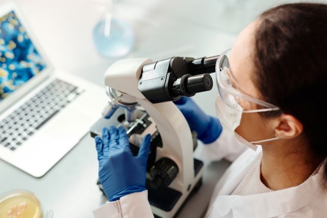
Cells do not function independently. Their behaviour is continuously influenced by the physical, chemical and biological conditions surrounding them. A cell receives signals from neighbouring cells, interacts with the extracellular matrix, responds to oxygen and nutrient availability, and adapts to local mechanical forces. Together, these influences form the tissue microenvironment, a complex and constantly changing environment that helps determine how cells grow, communicate, repair damage and respond to disease.
Traditional biological research often examines cells outside their original tissue context or studies individual components of the surrounding environment separately. Although these approaches provide valuable information, they may not fully reproduce the conditions that cells experience inside the human body. Artificial intelligence (AI), computational modelling and advanced imaging technologies are creating new possibilities for reconstructing these environments digitally. By combining molecular measurements, spatial information, tissue imaging and physical modelling, researchers can develop computational representations of the conditions surrounding cells.
This emerging field, known as computational tissue microenvironment reconstruction, could help scientists investigate how diseases begin, how abnormal cellular environments develop and why cells respond differently to apparently similar conditions. Its objective is not simply to create a digital image of tissue, but to build a scientifically useful model of the interactions that shape cellular behaviour.
What Makes Up the Tissue Microenvironment?
The tissue microenvironment is the local environment in which cells exist and interact. It includes both living components, such as neighbouring cells and immune populations, and non-living components, such as extracellular matrix proteins, dissolved nutrients, oxygen, chemical signals and physical structures. These elements work together to influence cellular identity, behaviour and function.
The extracellular matrix (ECM) is particularly important. It is a network of proteins and other molecules that provides structural support to tissues while also transmitting biochemical and mechanical information. Collagen, elastin, fibronectin and laminin are among the molecules that contribute to its organisation. Their distribution, density and physical properties influence how cells attach, move, divide and communicate. Changes in matrix composition or stiffness can alter cellular behaviour and contribute to processes such as fibrosis, tumour invasion and abnormal tissue repair.
The microenvironment also contains chemical gradients. Oxygen, glucose, growth factors, hormones and signalling molecules are not distributed uniformly throughout tissues. Cells located close to blood vessels may experience different nutrient and oxygen conditions from cells located deeper within a tissue. These variations can create distinct local environments even among cells that belong to the same tissue.
Mechanical conditions are equally relevant. Cells respond to stretching, compression, fluid movement and the stiffness of surrounding structures. These physical signals can influence gene expression and intracellular signalling through a process known as mechanotransduction. Therefore, understanding a cell’s biological behaviour requires more than identifying its molecular characteristics. Researchers must also understand the physical and chemical conditions in which that cell operates.
Why Reconstructing the Microenvironment Is Difficult
Conventional laboratory methods can isolate individual components of the tissue microenvironment, but reproducing their complete interactions is difficult. Cell cultures grown on flat laboratory surfaces, for example, do not necessarily experience the same spatial organisation, mechanical forces or molecular gradients found inside living tissues.
Even three-dimensional culture systems and organoids, which can reproduce selected aspects of tissue architecture, have limitations. Their internal regions may develop different nutrient and oxygen conditions, and their cellular composition may not fully match the original tissue. Vascular, immune and neural components can also be difficult to reproduce consistently.
Another challenge is tissue heterogeneity. A tissue sample may contain several cell types, each occupying a different location and experiencing a distinct set of signals. Two neighbouring cells may have different molecular states because they receive different signals from their surrounding environment. Traditional bulk molecular analysis can obscure these differences by combining measurements from many cells.
Spatial transcriptomics helps address part of this problem by measuring gene-expression patterns while retaining information about where those patterns occur within a tissue. Computational methods can then identify spatial regions, estimate cell-type distributions and investigate relationships between local molecular activity and tissue structure.
However, spatial measurements are still snapshots of complex biological systems. They may reveal where particular molecular patterns occur without fully explaining how the environment produced them or how they will change over time. Computational reconstruction attempts to connect these observations into a more complete representation.
How AI Reconstructs the Conditions Surrounding Cells
AI-based tissue microenvironment reconstruction begins by combining different types of biological and imaging data. These may include digital pathology images, spatial transcriptomics, spatial proteomics, single-cell sequencing, tissue morphology, extracellular matrix measurements and information about oxygen or nutrient availability.
Each data source describes a different aspect of the tissue. Histology images reveal structural organisation, spatial transcriptomics provides information about gene activity, and spatial proteomics helps identify the distribution of proteins. Single-cell datasets can provide reference profiles for identifying cell populations, while measurements of tissue mechanics can contribute information about stiffness and deformation.
AI models can integrate these datasets to estimate how cellular states relate to their surrounding conditions. For example, a model may identify a group of cells with a particular gene-expression pattern and examine whether those cells are located near blood vessels, immune-cell clusters or regions with altered extracellular matrix composition.
Deep-learning methods can also analyse high-resolution tissue images to identify cellular boundaries, tissue compartments and structural patterns. When these visual features are combined with molecular measurements, researchers can begin connecting tissue architecture with biological activity.
A 2025 review of AI approaches for cancer spatial omics describes how data-driven spatial AI, constraint-based approaches and mechanistic modelling can contribute to the interpretation of complex tissue environments. It also highlights the importance of model interpretability and integration with experimental research.
The result is not necessarily a perfect digital copy of the original tissue. It is a computational representation that estimates important relationships between cells, molecular signals and environmental conditions. Its scientific value depends on how accurately these relationships are supported by measurements and validated experiments.
Reconstructing Three-Dimensional Tissue Architecture
Most tissue samples used in molecular research are examined through two-dimensional sections. Although these sections provide valuable information, they cannot independently describe the complete three-dimensional arrangement of cells and structures. A blood vessel, for example, may appear as a small circular feature in one section but form part of an extended branching network across the surrounding tissue.
AI-assisted reconstruction can help connect information from multiple tissue sections to estimate three-dimensional organisation. Computer vision algorithms can identify corresponding structures, while computational registration methods align images collected from different positions. Molecular measurements can then be mapped onto the reconstructed architecture.
This process is important because the position of a cell can influence the signals it receives. Cells near a blood vessel may experience greater access to oxygen and nutrients, whereas cells farther away may exist in a relatively oxygen-poor environment. Similarly, cells located at the boundary between healthy and diseased tissue may interact with a different combination of immune cells and extracellular matrix components than cells located in the centre of the affected region.
Spatial omics research is increasingly moving towards integrating molecular mapping with three-dimensional tissue architecture and computational cell modelling. Recent reviews describe the limitations of relying only on isolated two-dimensional measurements and identify three-dimensional reconstruction as an important direction for more complete tissue analysis.
Modelling Oxygen, Nutrient and Chemical Gradients
The tissue microenvironment is not chemically uniform. Oxygen, glucose, metabolites and signalling molecules move through tissues according to factors such as blood supply, cellular consumption, diffusion distance and local tissue structure. These gradients can influence how cells behave and which biological pathways become active.
AI can help reconstruct these conditions by combining molecular measurements with mathematical models of transport and consumption. For example, researchers may use information about blood-vessel distribution, tissue density and cellular metabolic activity to estimate where oxygen availability is likely to be lower.
In cancer research, this can help investigate hypoxia, a condition in which cells experience insufficient oxygen availability. Hypoxic regions can develop when tumour growth exceeds the capacity of the local blood supply. These conditions may influence metabolism, angiogenesis, immune activity and treatment response.
Similar approaches could be used to investigate nutrient distribution in damaged tissues, metabolic changes during inflammation and chemical gradients around developing or regenerating structures. AI may identify relationships between the estimated local environment and the molecular states of nearby cells.
Nevertheless, predicted gradients should not be treated as direct measurements. Their accuracy depends on the quality of the underlying data and the assumptions used in the transport models. Experimental oxygen sensors, imaging techniques and metabolic measurements remain important for validating computational estimates.
Reconstructing the Extracellular Matrix and Mechanical Environment
The extracellular matrix is more than a passive support structure. Its composition, organisation and mechanical properties actively influence cellular behaviour. Changes in matrix stiffness, fibre alignment and molecular composition can alter how cells move, attach and respond to biochemical signals.
Computational reconstruction can combine tissue images, matrix measurements and mechanical modelling to estimate these properties. AI-based image analysis may identify collagen-rich regions, fibre orientation and structural differences between healthy and diseased tissue. Mathematical models can then examine how these structures may influence mechanical forces experienced by nearby cells.
This is particularly relevant to fibrosis, where excessive deposition of extracellular matrix components can make tissues progressively stiffer. In fibrotic tissue, altered mechanical conditions can influence the activity of fibroblasts and other cell populations, potentially reinforcing further matrix production.
Tumour tissues also demonstrate how mechanical changes can affect disease development. Abnormal matrix organisation and stiffness can influence tumour-cell movement, vascular function and interactions between cancer cells and surrounding immune populations. Research on tumour organoid modelling is increasingly examining how engineered matrices, microfluidic platforms and mechanical conditions can be used to reproduce selected features of the tumour environment.
AI-assisted reconstruction could help researchers compare the mechanical environments of healthy and diseased tissues, identify regions with unusual structural properties and investigate how these changes relate to cellular behaviour.
Understanding Cell-to-Cell Communication Within Reconstructed Tissues
Cells constantly exchange information through chemical signals, receptor interactions, direct contact and extracellular vesicles. These communication processes help coordinate tissue development, immune responses, metabolism and repair. However, the effects of a signal depend partly on which cells are located nearby and whether they possess the receptors and molecular machinery needed to respond.
Computational tissue reconstruction can help identify these relationships by mapping cell populations and their spatial arrangements. AI models can analyse the distribution of signalling molecules and receptors to estimate which neighbouring cell populations may communicate with one another.
For example, a reconstructed tissue model may reveal that immune cells are concentrated around a particular group of diseased cells. Researchers could then investigate whether the surrounding cells are producing signals that attract immune populations, suppress their activity or influence their movement. Similar analysis may reveal communication between fibroblasts, blood-vessel cells and epithelial cells during tissue repair.
Spatial analysis is especially valuable because the presence of two cell types in the same tissue does not prove that they interact directly. Their physical distance, molecular compatibility and local environment all influence whether communication is biologically plausible. AI can help prioritise potential interactions for further investigation, although computational predictions alone cannot establish that a signalling event has occurred.
Major Applications in Understanding Disease Development
Cancer Development and Tumour Microenvironment Analysis
Cancer is one of the most important applications of computational tissue microenvironment reconstruction. A tumour is not simply a collection of abnormal cells. It is a complex tissue ecosystem containing cancer cells, immune cells, fibroblasts, blood vessels and extracellular matrix components. Their interactions can influence tumour growth, invasion, immune evasion and responses to treatment.
AI can combine histology images with spatial molecular information to identify different regions within a tumour. Some regions may contain active immune populations, while others may show limited immune-cell access or altered metabolic conditions. Researchers can investigate whether these local differences are associated with particular tumour behaviours.
A 2025 review of spatial omics applications in cancer research describes how spatially resolved molecular measurements can reveal tumour heterogeneity, immune-cell infiltration patterns and interactions associated with disease progression and treatment resistance.
By reconstructing these environments computationally, researchers may gain a better understanding of why some tumour regions respond differently from others. This could support research into treatment resistance and the development of more context-sensitive experimental models.
Chronic Inflammation and Immune-Related Disorders
Inflammatory diseases involve complex interactions between immune cells, tissue-resident cells and local chemical signals. The environment surrounding immune cells can influence whether they become activated, migrate, release inflammatory molecules or enter regulatory states.
Computational reconstruction could help identify local inflammatory regions and examine how their molecular characteristics differ from nearby healthy tissue. AI may also investigate how immune-cell populations interact with fibroblasts, blood vessels and epithelial barriers.
This approach could be useful in studying conditions such as inflammatory bowel disease, rheumatoid arthritis and chronic skin inflammation. Researchers could compare tissue environments across disease stages to identify whether particular spatial patterns are associated with persistent inflammation or tissue damage.
Fibrosis and Abnormal Tissue Repair
Fibrosis develops when excessive connective tissue components accumulate and alter normal tissue structure. The surrounding environment becomes increasingly different from healthy tissue, with changes in matrix composition, stiffness, cellular activity and signalling.
AI-assisted reconstruction could help map these changes and identify regions where fibroblast activity, matrix deposition and mechanical alterations occur together. By comparing healthy and fibrotic tissue, researchers may identify local conditions associated with the development and persistence of abnormal tissue remodelling.
The ability to connect molecular activity with physical tissue properties is particularly important because fibrosis cannot be fully understood through gene expression alone. Its development also involves changes in the structure and mechanical behaviour of the surrounding tissue.
Neurodegenerative and Brain-Related Disorders
The brain has a highly specialised microenvironment involving neurons, astrocytes, microglia, blood vessels and extracellular structures. These components maintain communication, regulate nutrient delivery and support the conditions required for normal neural activity.
In neurodegenerative research, computational reconstruction could help investigate how local changes in immune activity, vascular function and cellular communication relate to neuronal damage. Researchers may also examine whether particular tissue regions show distinct molecular environments before or during the development of disease-associated changes.
Such models could contribute to the study of neuroinflammation, blood-brain barrier disruption and the relationship between neuronal and non-neuronal cells. However, the complexity of brain tissue means that reliable reconstruction requires high-quality spatial measurements and careful validation.
Connecting Computational Reconstruction With Organoids and Digital Tissue Models
Computational reconstruction becomes more useful when its predictions can be compared with experimental tissue models. Organoids, organ-on-chip systems and three-dimensional cell cultures offer opportunities to recreate selected features of human tissue in controlled laboratory conditions.
Organoids are three-dimensional structures developed from stem cells or tissue-derived cells. They can reproduce certain aspects of the organisation and function of real organs, although they do not automatically contain every cell type or environmental feature found in living tissue. Organ-on-chip systems use microfluidic technologies to control fluid movement, nutrient delivery and interactions between different cell populations.
AI can support the design and analysis of these experimental models. Researchers may use computational predictions to determine which cell populations, matrix properties or chemical conditions should be included in a laboratory model. Experimental observations can then be compared with the reconstructed tissue environment to assess whether the model reproduces the expected biological behaviour.
This creates an iterative research process. Computational models generate hypotheses, experimental systems test those hypotheses, and the resulting measurements help improve the models. Recent research on human model systems highlights the potential of combining engineered materials, organoids, microphysiological systems and computational modelling, while also recognising that no single platform currently reproduces the complete complexity of human biology.
Challenges and Limitations of AI-Based Tissue Reconstruction
Despite its potential, computational tissue microenvironment reconstruction faces substantial challenges. One major limitation is incomplete biological information. A tissue image may reveal cellular structure but provide limited information about molecular activity. Spatial transcriptomics may identify gene-expression patterns but not fully capture protein function, mechanical forces or rapid changes in chemical conditions.
Integrating different data types is also difficult. Imaging platforms, sequencing technologies and proteomic methods may operate at different resolutions and use different measurement standards. Aligning these datasets requires sophisticated computational methods and careful quality control.
Another challenge is the difference between correlation and causation. AI may identify a relationship between a particular cell population and a local molecular pattern, but this does not prove that the environment caused the cellular state. Both may be influenced by another biological process. Experimental interventions are necessary to test these proposed mechanisms.
Reconstructing dynamic tissue behaviour is even more demanding. Most available datasets represent a particular time point, whereas living tissues continuously change. Disease development may involve gradual shifts in oxygen availability, matrix structure, immune activity and cellular communication. A static computational model cannot fully explain these processes without longitudinal measurements or validated dynamic simulations.
There is also a risk of overestimating reconstruction accuracy. AI may fill gaps in incomplete datasets, but predicted molecular or spatial information is not equivalent to direct experimental measurement. Models must communicate uncertainty and be tested across independent tissue samples, laboratories and patient populations before their findings can be considered reliable.
The Future of Computational Tissue Microenvironment Reconstruction
The next stage of this field is likely to involve more integrated and dynamic tissue models. Advances in spatial transcriptomics, spatial proteomics, high-resolution microscopy and three-dimensional imaging are making it possible to collect increasingly detailed information about the organisation of biological tissues.
Multimodal AI models could combine these measurements with information about cellular behaviour, extracellular matrix composition and physical conditions. Instead of producing separate maps of gene expression, cell populations and tissue structure, they may develop unified representations that connect these features.
Another promising direction is the development of tissue digital twins. These are computational models designed to represent selected characteristics of a biological tissue and simulate how it may respond to changes in its environment. In research settings, such models could help investigate the potential effects of altered oxygen supply, inflammatory signals, matrix stiffness or experimental treatments.
However, a digital tissue model should not be confused with a complete virtual replica of a human organ. Its reliability depends on the biological processes represented, the quality of its data and the extent to which its predictions have been experimentally validated.
Future systems may also support patient-specific research by integrating molecular and spatial information from individual tissue samples. This could help explain why disease progression differs between individuals or why the same treatment produces different responses in different tissue environments. Achieving this goal will require standardised datasets, transparent algorithms, reproducible experimental methods and collaboration between computational scientists and biomedical researchers.
Conclusion
Computational tissue microenvironment reconstruction represents an important development in the study of disease because it focuses on the conditions surrounding cells rather than examining cellular activity in isolation. The extracellular matrix, neighbouring cells, oxygen availability, nutrients, chemical signals and mechanical forces all influence how tissues function and how diseases develop.
AI can help bring these different dimensions together by integrating spatial molecular measurements, digital pathology, three-dimensional imaging and computational modelling. These approaches may allow researchers to identify disease-associated tissue regions, investigate abnormal cellular communication and understand how local environmental changes influence biological behaviour.
Although current methods cannot reproduce every feature of living human tissue, they provide increasingly sophisticated ways to investigate its complexity. As spatial technologies, experimental tissue models and AI methods advance, computational reconstruction may become a valuable tool for understanding disease development, improving laboratory models and supporting more precise biomedical research.
You may be interested
Wellness Decision Overload Architecture: Conflicting health information changes
Anshika Jain - November 18, 2026Access to health information has expanded significantly through digital platforms, healthcare applications, social media, online communities and search engines. Individuals can now explore information about nutrition, exercise,…

Health Behaviour Switching Costs: Mental effort replace unhealthy routines
Anshika Jain - November 18, 2026Changing an unhealthy habit is often more complicated than simply understanding why it should be changed. People may recognise the importance of regular exercise, balanced nutrition, adequate…

Physiological Self-Regulation Capacity: Individuals Maintain Biological Stability
Anshika Jain - November 18, 2026The human body continuously responds to changes in its internal and external environment. Everyday activities such as walking, working, exercising, eating, sleeping, experiencing emotional pressure and adapting…
Most from this category

Food Fermentation Metabolite Competition: Microorganisms compete to produce compounds
Anshika Jain - November 18, 2026
Plant Pigment Stability Prediction: Cooking and storage influence anthocyanins
Anshika Jain - November 18, 2026
Starch Retrogradation Intelligence:Cooling and reheating change properties of cooked starch
Anshika Jain - November 18, 2026
Biological Response Reproducibility Intelligence: Same health intervention produces inconsistent results
Anshika Jain - November 17, 2026
Food Fermentation Metabolite Competition: Microorganisms compete to produce compounds
Anshika Jain - November 18, 2026Plant Pigment Stability Prediction: Cooking and storage influence anthocyanins
Anshika Jain - November 18, 2026Starch Retrogradation Intelligence:Cooling and reheating change properties of cooked starch
Anshika Jain - November 18, 2026




.png)
Leave a Comment
You must be logged in to post a comment.