
Biological Tissue Aging Maps: AI Distinguish Functional Aging From Changes in Appearance

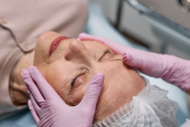
Aging is often recognised through visible changes. Wrinkles become more pronounced, skin loses elasticity, hair changes colour, facial contours shift and posture may gradually become different. These signs are commonly used to describe how a person is aging, but they reveal only part of a much more complicated biological process. Beneath the visible surface, tissues undergo changes in cellular activity, structural organisation, repair capacity, metabolism and physiological performance. These changes do not necessarily occur at the same speed or follow the same pattern across the body.
A person may have visible signs of aging while maintaining relatively strong physical function, whereas another individual may appear younger but experience reduced muscle performance, declining organ function or changes in tissue repair. This difference raises an important scientific question: can biological aging be measured independently of appearance, and can artificial intelligence help researchers understand how different tissues age at different rates?
Biological Tissue Aging Maps is an emerging concept that describes the use of artificial intelligence to analyse tissue-specific structural, molecular and functional information and represent how aging develops across different parts of the body. Instead of assigning a single biological age to an entire person, such a system could examine the skin, muscles, blood vessels, brain, lungs, kidneys and other tissues separately. It could compare visible changes with deeper biological indicators to investigate whether a tissue is primarily showing an alteration in appearance, a reduction in function or a combination of both.
AI could contribute by analysing medical images, tissue samples, molecular profiles, clinical measurements and longitudinal health records. Its ability to recognise complex patterns may help researchers distinguish cosmetic or structural aging from changes that affect physiological performance.
Understanding the Difference Between Visible Aging and Functional Aging
Visible aging refers to changes that can be observed externally or through imaging. In the skin, these may include wrinkles, pigmentation changes, reduced elasticity, changes in thickness and alterations in facial volume. These characteristics are influenced by intrinsic aging as well as environmental exposures such as ultraviolet radiation, smoking, pollution, nutrition and lifestyle. Although appearance can provide useful information about tissue structure, it cannot independently reveal the complete biological condition of that tissue.
Functional aging concerns changes in what a tissue or organ can do. Muscle tissue, for example, may gradually lose strength, power and regenerative capacity. Blood vessels may become stiffer, affecting their ability to expand and regulate circulation. The lungs may experience changes in elasticity and respiratory performance, while the kidneys may show reductions in filtration capacity. These changes can develop without producing obvious external signs.
The distinction is important because structural appearance and physiological performance are related but not interchangeable. A skin image may reveal wrinkles and texture changes, but it cannot directly establish the skin’s wound-healing capacity, immune activity or cellular senescence. Similarly, the visible appearance of a person’s face cannot reliably determine the biological age of the heart, brain or kidneys.
Biological aging is also asynchronous. Different organs and tissues may age at different rates within the same individual. Genetic characteristics, blood supply, metabolic demands, environmental exposure, inflammation and previous injuries all contribute to this variation. Consequently, a single age estimate may conceal important differences between individual tissues.
What Are Biological Tissue Aging Maps?
Biological Tissue Aging Maps can be understood as integrated representations of age-related changes across specific tissues and organ systems. Their purpose would be to connect different forms of biological evidence rather than relying on one visible feature or one molecular measurement.
For example, a tissue map for the skin might combine microscopic images, measurements of collagen organisation, gene-expression patterns, inflammatory markers and indicators of barrier function. A muscle-focused map could examine muscle size, tissue composition, strength measurements, movement performance and molecular markers associated with repair and cellular maintenance. A blood-vessel map might combine vascular imaging, arterial stiffness measurements and molecular indicators of inflammation or endothelial function.
AI could analyse these inputs to identify patterns associated with aging and compare them with reference populations. The resulting representation might show that one tissue displays relatively advanced structural changes while another maintains better functional characteristics. It could also reveal differences between visible appearance and molecular or physiological measurements.
The concept should not be confused with a single established medical test. At present, researchers use different biological aging clocks, imaging methods and tissue-specific measurements, each designed for particular purposes. A comprehensive map combining all these dimensions remains a research ambition rather than a routine clinical assessment.
How AI Can Analyse Tissue-Specific Aging
Medical Imaging and Histological Analysis
Medical imaging provides an important source of information about tissue structure. Magnetic resonance imaging can reveal changes in the brain, muscles and other soft tissues, while ultrasound and specialised imaging techniques can examine selected structural and functional characteristics. Histopathology provides another level of detail through microscopic examination of tissue samples.
AI models can analyse medical images to identify patterns that may be difficult to quantify consistently through manual assessment. Deep-learning systems can examine tissue architecture, cell distribution, structural organisation and other visual characteristics. When trained on appropriately labelled datasets, these models may estimate age-related patterns and compare them across individuals.
A particularly important development was reported in a 2026 Nature Medicine study that analysed 25,712 whole-slide histopathological images from 40 tissue types across 983 individuals. Researchers developed deep-learning-based tissue clocks that estimated biological age from microscopic tissue structure. The study reported associations between tissue-specific age estimates and indicators including telomere attrition, subclinical pathology and comorbidities. It also explored whether tissue-specific age gaps could be estimated from blood-based information by integrating histological and transcriptomic data. This research demonstrates how AI can investigate aging beyond visible facial appearance, although further validation is necessary before such methods can be used as routine clinical measurements.
Molecular and Epigenetic Aging Signals
Tissue aging is also reflected in molecular changes that cannot be identified through ordinary photographs. DNA methylation, gene expression, protein abundance and metabolic activity can provide information about changes occurring within cells and tissues. These measurements are often described as omics data because they capture large collections of biological features.
Epigenetic clocks are among the best-known examples of molecular aging models. They use patterns of DNA methylation to estimate age or assess aspects of biological aging. Different clocks have been developed for different research objectives, including chronological-age estimation, health-related risk assessment and the pace of aging. However, a clock trained to predict chronological age does not necessarily measure tissue function or predict how well an organ performs.
AI can help integrate epigenetic information with transcriptomic, proteomic and metabolomic data. This may allow researchers to investigate whether molecular patterns are associated with tissue-specific structural changes or functional decline. A 2025 review of deep aging clocks describes the use of AI across epigenetics, transcriptomics, metabolomics, microbiome information and imaging-based approaches. It also highlights the potential of deep-learning methods to capture complex, non-linear relationships that may be missed by simpler models.
Functional Measurements and Physiological Performance
To distinguish functional aging from changes in appearance, AI systems must include information about what tissues actually do. Muscle aging, for example, cannot be understood through muscle volume alone. Strength, power, endurance, coordination and recovery after physical activity are also relevant. Similarly, the condition of the cardiovascular system requires measurements that reflect circulation and vascular performance, not simply images of blood vessels.
Researchers could combine these measurements with molecular and imaging information to investigate whether visible structural changes correspond to meaningful functional differences. For example, a model might compare muscle imaging with grip strength, walking performance and indicators of muscle metabolism. It could then examine whether a particular structural pattern is consistently associated with reduced physical performance.
This combination is important because a tissue may show age-related changes without experiencing an equivalent reduction in function. Conversely, functional limitations may appear before major structural differences become visible. AI could help identify these mismatches and investigate the biological mechanisms associated with them.
Why Different Tissues Need Different Aging Maps
Aging does not follow an identical biological pathway throughout the body. Each tissue has its own cellular composition, metabolic demands, regenerative capacity and exposure to environmental stress. A useful tissue-aging framework must therefore recognise organ-specific characteristics instead of applying one universal aging score to every part of the body.
Skin aging is influenced by changes in collagen, elastin, cellular turnover, pigmentation and barrier function. Ultraviolet exposure can accelerate certain structural changes, meaning that visible skin aging may reflect both intrinsic biological processes and accumulated environmental damage. A skin-focused AI model could compare photographs with molecular or tissue-level measurements to investigate whether appearance accurately reflects deeper tissue condition.
Muscle aging involves changes in muscle fibres, strength, mitochondrial function, protein turnover and the interaction between muscle and the nervous system. A person may maintain a relatively youthful appearance while experiencing reduced muscle strength or physical endurance. An AI system that integrates imaging, movement measurements and molecular indicators could help researchers examine these differences.
In the cardiovascular system, aging may involve arterial stiffening, endothelial changes and alterations in the structure of blood vessels. These processes can affect physiological performance without producing visible external signs. Similarly, brain aging may involve changes in neural networks, tissue volume, white-matter integrity and cognitive performance. Brain imaging combined with cognitive assessments and molecular data may help researchers distinguish structural age estimates from functional changes.
The kidney and lung provide further examples. Changes in filtration, respiratory capacity and tissue structure may develop at different rates among individuals. A general biological age score may not adequately represent these organ-specific trajectories. Tissue-specific maps could therefore provide a more detailed research framework for understanding why two people of the same chronological age may have different patterns of physiological aging.
Online Internship with Certificate
You may be interested
Gut Virome Intelligence: Infect Bacteria Influence the Human Gut Ecosystem
Anshika Jain - November 6, 2026The human gut is home to a complex ecosystem containing bacteria, fungi, archaea and viruses. While gut bacteria have received considerable attention in research on digestion, immunity…
Human–Environment Health Interaction Maps: AI Connect Indoor Lighting
Anshika Jain - November 6, 2026Human health is influenced not only by genetics, nutrition, physical activity and medical care but also by the environments in which people spend their everyday lives. Homes,…
AI Predict Which Preventive Health Interventions Will Remain Effective
Anshika Jain - November 6, 2026Preventive healthcare traditionally focuses on identifying risk factors and recommending interventions that can reduce the likelihood of disease. However, a life-course approach recognises that health is shaped…
Most from this category
Science of Medical Device Fatigue: People Stop Using Health Technologies
Anshika Jain - November 6, 2026
AI Model How Food Packaging Chemicals Migrate Into Different Types of Food
Anshika Jain - November 6, 2026
Retinal Microvascular Intelligence: Early Signs of Changes Beyond Eye Health
Anshika Jain - November 5, 2026
Hair Follicle Biology AI: Machine Learning Distinguish Nutritional Factors
Anshika Jain - November 5, 2026
Science of Medical Device Fatigue: People Stop Using Health Technologies
Anshika Jain - November 6, 2026AI Model How Food Packaging Chemicals Migrate Into Different Types of Food
Anshika Jain - November 6, 2026Retinal Microvascular Intelligence: Early Signs of Changes Beyond Eye Health
Anshika Jain - November 5, 2026


.png)
Leave a Comment
You must be logged in to post a comment.