
Extracellular Vesicle Communication Analysis: Tiny particles released by cells transport biological information between tissues

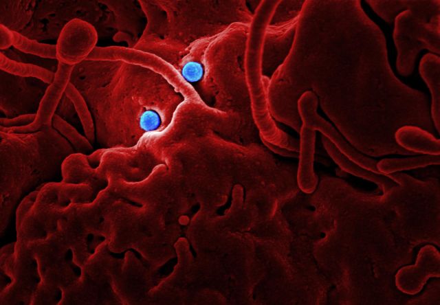
The human body contains billions of cells that continuously exchange information to coordinate biological activities. Cells communicate through chemical signals, hormones, electrical impulses and direct physical interactions. However, another important communication mechanism operates through extremely small membrane-enclosed particles released into the surrounding environment. These particles, known as extracellular vesicles (EVs), carry biological materials from one cell to another and can influence the behaviour of distant or neighbouring tissues.
Extracellular vesicles contain different combinations of proteins, lipids, nucleic acids and other molecular components. Their contents can reflect the condition of the cells that release them. After entering extracellular spaces or bodily fluids, these particles may interact with recipient cells and deliver molecular information that influences cellular activity. This communication is relevant to normal processes such as immune regulation, tissue repair and blood vessel formation, as well as disease-related processes including cancer progression, inflammation and neurological disorders.
Despite their small size, extracellular vesicles represent a highly complex biological communication system. Their molecular contents vary according to their cellular origin, physiological environment and release mechanisms. Understanding which cell produces a particular vesicle, what information it carries, where it travels and how recipient cells respond requires the integration of several types of biological data.
Artificial intelligence could help researchers investigate this complexity. By analysing molecular profiles, microscopy images, sequencing results and cellular interaction networks, AI models may identify patterns that explain how extracellular vesicles participate in communication between tissues. This emerging research direction, called Extracellular Vesicle Communication Analysis, could contribute to a deeper understanding of cell-to-cell signalling, disease mechanisms and the development of future diagnostic technologies.
Understanding Extracellular Vesicles and Their Biological Functions
Extracellular vesicles are membrane-enclosed particles released by cells into the extracellular environment. Their lipid bilayer protects and carries biological molecules that may participate in communication with other cells. They are found in several biological fluids, including blood, urine, saliva, breast milk and cerebrospinal fluid, making them particularly interesting for biomedical research.
Extracellular vesicles are not all identical. They differ in size, formation pathways, molecular composition and biological functions. Exosomes are generally associated with the endosomal pathway, whereas microvesicles are formed through outward budding of the plasma membrane. However, distinguishing these categories in experimental samples can be difficult. For this reason, current research recommendations encourage the use of operational descriptions such as small or large extracellular vesicles unless their specific biological origin has been demonstrated.
The molecular cargo of extracellular vesicles can include messenger RNA, microRNA, other non-coding RNAs, DNA fragments, membrane proteins, enzymes, lipids and signalling molecules. These components may influence the activity of recipient cells by modifying gene expression, activating signalling pathways or changing metabolic behaviour.
For example, an immune cell may release extracellular vesicles containing molecules that influence the activity of other immune cells. Cells involved in tissue repair may release vesicles that participate in communication with surrounding cells. Similarly, tumour cells can release vesicles containing molecular signals that influence their surrounding environment.
The importance of extracellular vesicles lies not simply in their ability to transport molecules, but in the biological information represented by their combined molecular contents.
How Extracellular Vesicles Transport Biological Information Between Tissues
Extracellular vesicle communication involves several interconnected stages. First, a cell produces and releases vesicles containing selected molecular components. These vesicles enter the surrounding extracellular environment, where they may interact with neighbouring cells, travel through biological fluids or become captured by recipient tissues.
Once a vesicle reaches a potential recipient cell, it may interact with the cell surface through receptor-mediated binding or other molecular recognition mechanisms. Some vesicles are internalised through processes such as endocytosis, while others may deliver their contents through membrane fusion or related mechanisms. The precise route depends on the vesicle, recipient cell and surrounding biological conditions.
After uptake or interaction, vesicle-associated molecules may influence the recipient cell. For instance, transferred RNA can potentially affect gene expression, while proteins and lipids may alter signalling pathways or membrane-related activities. However, detecting a molecule inside an extracellular vesicle does not automatically prove that it will produce a functional change in another cell. Researchers must establish whether the cargo is delivered, remains biologically active and reaches a sufficient concentration to influence the recipient.
This communication can occur locally or over longer distances. Vesicles released into the bloodstream may encounter cells in different organs, although their distribution is influenced by circulation, clearance mechanisms, surface properties and interactions with biological barriers. Understanding these factors is essential for determining whether an extracellular vesicle can genuinely act as a messenger between distant tissues.
Why Extracellular Vesicle Communication Is Difficult to Decode
The main challenge in studying extracellular vesicles is their substantial biological heterogeneity. A single biological sample may contain vesicles released by different cell types, each carrying distinct combinations of proteins, lipids and nucleic acids. Even vesicles produced by the same cell population may differ according to the cell’s metabolic state, exposure to stress and surrounding environment.
This variation makes it difficult to identify the precise origin and function of individual vesicles. A sample collected from blood, for example, may contain vesicles associated with immune cells, platelets, endothelial cells and other tissues. Their molecular contents can overlap, which complicates the identification of reliable cell-specific signatures.
Another challenge is separating extracellular vesicles from other particles present in biological fluids. Lipoproteins, protein aggregates and other non-vesicular particles can be isolated alongside vesicles and may interfere with molecular measurements. The International Society for Extracellular Vesicles addresses these issues through its MISEV2023 recommendations, which emphasise appropriate separation, characterisation, controls and transparent reporting. The guidelines also explain why no single measurement is sufficient to characterise every aspect of an extracellular vesicle preparation.
The biological effects of vesicles introduce further complexity. A vesicle’s cargo does not necessarily determine its function independently. Its effect may depend on the recipient cell’s receptors, internal signalling networks, metabolic condition and ability to process the transferred material. Therefore, researchers must study both the molecular information carried by vesicles and the biological context in which that information is received.
These challenges create an opportunity for AI systems that can analyse multiple biological variables together rather than treating extracellular vesicles as a uniform population.
How Artificial Intelligence Can Decode Extracellular Vesicle Communication
Identifying the Cellular Origin of Extracellular Vesicles
Understanding where a vesicle originates is essential for interpreting its biological significance. Different cell types produce extracellular vesicles with different molecular characteristics, although many markers are shared across populations.
Machine learning models can examine combinations of surface proteins, RNA profiles, lipid composition and other molecular measurements to estimate the likely cellular origin of vesicles. Instead of relying on one marker, a model can analyse multiple features and identify patterns associated with particular cell populations.
For example, researchers may compare vesicles collected from healthy and inflamed tissues to investigate whether their molecular signatures indicate different cellular sources. AI could also help distinguish changes in vesicle composition from changes in the proportions of vesicles released by different cell types.
Single-vesicle analysis is particularly valuable for this purpose. Conventional bulk analysis measures the combined molecular contents of many vesicles, potentially hiding rare populations with distinctive biological characteristics. Newer single-vesicle technologies can examine individual particles and reveal differences that are not visible in population-level averages. Research published in 2024 and 2025 has highlighted the growing importance of single-EV analysis for identifying molecular diversity and improving the interpretation of extracellular vesicle populations.
Analysing RNA, Protein and Lipid Cargo
Extracellular vesicles transport different classes of biological molecules, and each provides a different type of information. RNA cargo may influence gene expression in recipient cells, proteins may participate in signalling or molecular recognition, and lipids can affect membrane properties and cellular interactions.
AI models can integrate these molecular measurements to identify coordinated cargo patterns. For example, a vesicle population may contain particular microRNAs alongside proteins associated with inflammation. Analysing these components together may provide a more informative biological signature than studying either RNA or protein data independently.
Deep learning can also support the interpretation of complex sequencing and mass spectrometry datasets. Algorithms may identify molecular combinations associated with particular cellular conditions, distinguish disease-associated patterns and help prioritise candidate cargo molecules for experimental investigation.
The challenge is to determine whether these molecular signatures represent actual biological communication. A detected RNA molecule may be present in a vesicle without being delivered to a recipient cell in a functional quantity. AI predictions therefore need to be connected with uptake experiments, molecular measurements and functional studies.
Predicting Which Tissues May Receive Vesicle Signals
Extracellular vesicles do not interact equally with every cell they encounter. Their surface molecules, membrane composition and biological environment can influence their distribution and interactions with recipient tissues.
AI could help investigate these relationships by combining vesicle surface profiles with information about receptor expression in potential recipient cells. Computational models may identify candidate interactions between vesicle-associated proteins and cell-surface receptors, helping researchers prioritise possible communication pathways for laboratory testing.
For instance, researchers could investigate whether vesicles released by a particular cell population contain surface proteins associated with interactions with endothelial cells or immune cells. Models could compare these signatures with receptor profiles from different tissues and estimate which interactions deserve further investigation.
Such predictions would not establish the actual destination of vesicles in a living organism. Distribution depends on many additional factors, including blood flow, tissue barriers, clearance by immune cells and the physical properties of vesicles. Nevertheless, computational analysis could narrow the range of possible interactions and guide more focused experiments.
Reconstructing Cell-to-Cell Communication Networks
A particularly promising application is the reconstruction of cellular communication networks. Instead of examining one vesicle or one cell interaction, researchers could model how multiple cell populations exchange vesicle-associated signals across a tissue or organ system.
Graph-based machine learning may be useful for this purpose. In a communication network, individual cell populations can be represented as nodes, while potential vesicle-mediated interactions form connections between them. Each connection can include information about the vesicle’s molecular cargo, the suspected recipient cell and the experimental evidence supporting the interaction.
Researchers could use these networks to investigate how changes in one cell population influence other parts of a biological system. For example, inflammation in one tissue might alter the composition of released vesicles, which could then influence immune or vascular cell activity elsewhere.
A systems-biology approach can also combine vesicle data with gene-expression profiles, receptor measurements and tissue-specific information. A 2025 review of extracellular vesicle systems biology and multi-omics research describes how computational integration is helping connect vesicle composition with cellular origin, disease-associated molecular patterns and potential biological functions.
The Role of AI in Understanding Extracellular Vesicles in Disease
Cancer Progression and Communication With Surrounding Tissues
Cancer cells release extracellular vesicles that can participate in communication with surrounding cells. Their cargo may influence immune activity, blood vessel formation, tissue remodelling and interactions between tumour cells and their microenvironment.
Some tumour-associated vesicles carry molecules that may contribute to changes in immune-cell behaviour. Others may participate in signalling between cancer cells and stromal cells, which form part of the supportive tissue surrounding a tumour. These interactions can influence how the tumour environment develops. AI models could analyse vesicle-associated RNA, proteins and lipids from cancer samples to identify molecular signatures associated with particular tumour characteristics.
Such analysis could also contribute to liquid biopsy research. Since extracellular vesicles are present in blood and other biological fluids, researchers are investigating whether their molecular contents can provide information about diseases without requiring repeated tissue biopsies. However, clinical applications require reliable evidence that candidate signals are sufficiently specific, reproducible and useful across independent patient groups.
Brain Communication and Neurological Disorders
The brain contains specialised cell populations, including neurons, astrocytes, microglia and oligodendrocytes. These cells communicate through several mechanisms, and extracellular vesicles are being investigated as one possible contributor to molecular exchange within nervous tissue.
Vesicles released by neural cells may contain proteins and RNA molecules associated with neuronal activity, inflammation and cellular stress. Researchers are also investigating how extracellular vesicles may participate in communication between the central nervous system and other parts of the body.
AI could help analyse complex vesicle profiles from cerebrospinal fluid and blood, potentially identifying patterns associated with neurological conditions. By integrating molecular measurements with clinical and experimental data, computational models may help researchers investigate disease-associated changes in cell communication.
However, the presence of a particular vesicle signature in blood does not automatically establish that it originated in the brain. Identifying tissue origin and demonstrating the biological pathway involved remain important research challenges.
Inflammation and Immune-System Regulation
Immune cells release extracellular vesicles that may influence the activity of other immune cells and surrounding tissues. Their molecular cargo can reflect whether the originating cells are responding to infection, tissue damage or inflammatory signals.
AI-based communication analysis could help identify how vesicle composition changes during different immune responses. Researchers may compare molecular signatures from resting and activated immune cells and examine whether particular vesicle populations are associated with changes in inflammatory signalling.
This could improve understanding of how local immune responses influence distant tissues. It may also help identify candidate molecular markers for studying inflammatory diseases. Nevertheless, the effects of extracellular vesicles depend on the recipient cells and the wider biological environment, so a predicted communication network requires functional validation.
AI Technologies Supporting Advanced Extracellular Vesicle Research
The development of AI-based extracellular vesicle analysis depends on several complementary technologies. Deep learning is useful for analysing microscopy images, identifying particle characteristics and classifying molecular patterns. Convolutional neural networks can support automated image interpretation, while other machine learning approaches can classify vesicle populations using protein, RNA and lipid measurements.
Graph neural networks may help reconstruct communication networks by representing relationships between vesicles, originating cells and recipient tissues. These systems can analyse complex interactions in which one cell population communicates with several other populations through different molecular pathways.
Multi-omics integration is another important direction. By combining transcriptomics, proteomics, lipidomics and metabolomics, researchers can investigate extracellular vesicles as complex biological information carriers rather than isolated collections of molecules. A 2026 review of machine learning in extracellular vesicle research describes applications involving automated imaging, multi-omics integration, disease classification, therapeutic engineering and quality control. It also identifies data sparsity, batch variability and model explainability as important technical challenges.
Microfluidic technologies may further improve this research by enabling the controlled separation, capture and analysis of small quantities of extracellular vesicles. When combined with machine learning, these platforms could support more efficient processing of experimental samples and potentially enable faster molecular profiling.
The combination of these technologies may eventually allow researchers to develop integrated systems that connect vesicle identity, molecular cargo, cellular origin and recipient-cell response.
Challenges in Developing Reliable AI-Based Communication Models
Despite significant progress, AI-based extracellular vesicle research faces several limitations. One of the most important is the lack of standardised experimental data. Differences in sample collection, storage, isolation procedures and analytical techniques can influence the measured characteristics of vesicles. Models trained on data from one laboratory may therefore perform differently when applied to another dataset.
Another difficulty is the identification of rare vesicle populations. A small number of biologically important particles may be hidden within a much larger and more heterogeneous population. Single-vesicle technologies can help address this problem, but their measurements may still be affected by detection limits, technical noise and differences in instrument sensitivity.
The distinction between vesicle-associated molecules and contaminants is also essential. Some proteins and nucleic acids detected in an isolated sample may originate from non-vesicular particles rather than extracellular vesicles. If such contamination is not adequately controlled, an AI model may learn misleading molecular patterns.
Predicting communication is also more complicated than identifying molecular similarities. A vesicle may contain a particular protein, but that does not prove it will reach a specific tissue or activate a biological pathway. Researchers must demonstrate the complete sequence of events, including vesicle release, transport, recipient-cell interaction and functional response.
Finally, AI models must be interpretable and independently validated. Researchers need to understand which molecular features contribute to a prediction and whether those features correspond to established biological mechanisms. Computational predictions should guide experimental investigation rather than replace it.
The Future of Extracellular Vesicle Communication Analysis
Future research may move towards personalised and tissue-specific extracellular vesicle communication models. Instead of analysing average vesicle profiles from large cell populations, scientists could investigate how individual vesicles differ and how these differences relate to particular cell states.
Single-cell and single-vesicle technologies may make it possible to examine communication at increasingly detailed levels. AI could help connect an individual cell’s molecular characteristics with the vesicles it releases and the responses observed in potential recipient cells. This may reveal communication patterns that remain hidden in conventional bulk measurements.
Another promising direction is the development of digital representations of tissue communication networks. Such models could integrate information about cell populations, extracellular vesicle cargo, receptor availability and tissue environments. Researchers could then simulate possible changes in communication under different experimental conditions and identify the pathways that deserve further study.
AI may also support the development of extracellular vesicle-based therapeutics. Researchers are investigating the possibility of using engineered vesicles to deliver selected biological materials to specific cells. Computational systems could help examine cargo selection, surface properties and potential recipient-cell interactions.
The field is also moving towards stronger research standardisation. More consistent experimental procedures, transparent reporting and independent validation will be essential for translating computational findings into meaningful biological discoveries. The MISEV recommendations provide an important foundation for improving the reliability and reproducibility of extracellular vesicle studies.
Conclusion
Extracellular vesicles provide cells with an important mechanism for exchanging biological information. By carrying proteins, lipids, RNA and other molecular components, they may influence cellular activity, immune regulation, tissue repair and disease-related processes. Their communication networks are highly complex because vesicle composition, cellular origin, transport behaviour and recipient-cell responses can vary considerably.
Artificial intelligence offers promising methods for investigating this complexity. Machine learning can identify molecular signatures, deep learning can analyse microscopy and sequencing data, and graph-based approaches can help reconstruct potential communication networks between cells and tissues. Integrating these methods with single-vesicle analysis and multi-omics research may provide a more detailed understanding of how extracellular vesicles participate in biological signalling.
However, decoding vesicle communication requires more than identifying molecular patterns. Researchers must establish where vesicles originate, how they reach recipient cells and whether their cargo produces measurable biological effects. Experimental validation, reliable data and standardised methodologies will remain essential.
Ultimately, Extracellular Vesicle Communication Analysis could become an important research direction for understanding how tissues exchange information at the molecular level. By combining AI with advanced cellular and molecular technologies, scientists may uncover previously hidden communication pathways and improve research into disease biomarkers, tissue interactions and future therapeutic strategies.
Online Internship with Certificate
You may be interested

Glycoprotein Structure Prediction: Changes in Protein-linked sugar structures
Anshika Jain - November 19, 2026Proteins are responsible for many essential biological activities, including cellular communication, immune defence, molecular transport, tissue development and the regulation of biochemical reactions. However, proteins do not…

Cellular Autophagy Regulation Intelligence: Cells recycle damaged components
Anshika Jain - November 19, 2026Every living cell continuously performs a complex maintenance process to preserve its internal environment, remove damaged structures and maintain normal biological activity. Proteins become misfolded, cellular membranes…

RNA Modification Mapping: Chemical Modifications to RNA Influence
Anshika Jain - November 19, 2026Gene expression is often described as the process through which information stored in DNA is used to produce functional RNA and proteins. However, the activity of genes…
Most from this category
Organelle-Level Disease Intelligence: Dysfunction in Cellular Structures
Anshika Jain - November 19, 2026
Behavioural Adaptation Under Uncertainty: People maintain health routines
Anshika Jain - November 19, 2026
Wellness Decision Overload Architecture: Conflicting health information changes
Anshika Jain - November 18, 2026
Health Behaviour Switching Costs: Mental effort replace unhealthy routines
Anshika Jain - November 18, 2026
Organelle-Level Disease Intelligence: Dysfunction in Cellular Structures
Anshika Jain - November 19, 2026Behavioural Adaptation Under Uncertainty: People maintain health routines
Anshika Jain - November 19, 2026Wellness Decision Overload Architecture: Conflicting health information changes
Anshika Jain - November 18, 2026

RNA Modification Mapping: Chemical Modifications to RNA Influence
November 19, 2026

.png)
Leave a Comment
You must be logged in to post a comment.