
Organelle-Level Disease Intelligence: Dysfunction in Cellular Structures

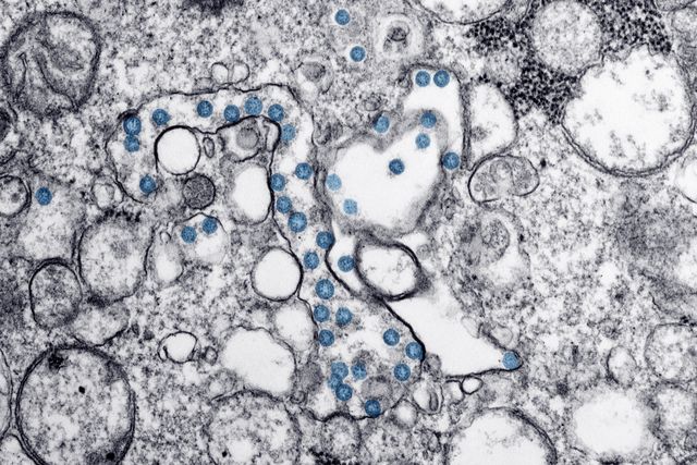
Many complex diseases develop through biological changes that begin long before their symptoms become noticeable. Some of these changes occur inside individual cells, where specialised structures known as organelles perform essential functions such as producing energy, processing proteins, regulating calcium, managing cellular waste and coordinating internal communication. When these structures become damaged or stop functioning correctly, the consequences can extend across entire cells, tissues and organs. Such disruptions are associated with several conditions, including neurodegenerative disorders, metabolic diseases, cardiovascular conditions and certain cancers.
Understanding these processes is challenging because organelles do not operate independently. Mitochondria influence energy production and cellular stress responses, the endoplasmic reticulum helps manufacture and fold proteins, lysosomes break down unwanted materials, and the Golgi apparatus modifies and transports molecules. Their functions are interconnected, meaning that dysfunction in one structure can affect several others. A disturbance in mitochondrial activity, for example, may influence calcium regulation, cellular signalling and the mechanisms responsible for removing damaged components.
Organelle-level disease intelligence explores how artificial intelligence can help researchers investigate these relationships. By analysing microscopy images, molecular measurements, genomic information and other biological datasets, AI may identify patterns that are difficult to recognise through conventional analysis alone. The objective is to understand how cellular structures contribute to disease development, how dysfunction spreads through biological systems and whether these findings can support improved disease classification and therapeutic research.
This emerging research direction could strengthen the connection between cellular biology and disease investigation. Rather than studying individual organelles as isolated structures, researchers can use computational methods to examine their interactions as part of a larger biological network. However, its success depends on reliable experimental evidence, biological interpretation and careful validation rather than computational predictions alone.
Understanding the Role of Organelles in Cellular Health
Organelles are specialised structures within cells that perform different but closely coordinated activities. Their functions are essential for maintaining cellular balance, responding to environmental changes and supporting the activities of tissues and organs. Although each organelle has particular responsibilities, the overall health of a cell depends on how effectively these structures communicate and coordinate their processes.
Mitochondria are commonly recognised as the energy-producing structures of cells because they generate much of the energy required for cellular activity through oxidative phosphorylation. However, their functions extend beyond energy production. They also participate in calcium regulation, metabolic signalling, cellular stress responses and programmed cell death. When mitochondrial function becomes impaired, cells may experience energy shortages, increased oxidative stress and changes in signalling pathways.
The endoplasmic reticulum performs another important role by supporting protein synthesis, folding and processing. It also contributes to lipid metabolism and calcium storage. When proteins fail to fold correctly or the endoplasmic reticulum experiences prolonged stress, cellular processes can become disrupted. Lysosomes, meanwhile, are responsible for breaking down unwanted materials and recycling damaged cellular components. If their degradation systems become inefficient, waste products may accumulate and interfere with normal cellular function.
The Golgi apparatus, peroxisomes, lipid droplets and other cellular structures also contribute to maintaining internal balance. These organelles participate in protein transport, lipid processing, detoxification and metabolic regulation. Their combined activity demonstrates why cellular health cannot be fully understood by examining only one structure at a time.
How Organelle Dysfunction Contributes to Complex Diseases
Mitochondrial Dysfunction and Neurodegenerative Conditions
Mitochondrial dysfunction is an important area of investigation in neurodegenerative diseases because nerve cells require substantial amounts of energy to maintain their structure, communicate with other cells and perform specialised functions. Disruptions in mitochondrial activity may affect energy availability, calcium regulation, oxidative balance and cellular survival.
In conditions such as Alzheimer’s and Parkinson’s disease, researchers investigate mitochondrial abnormalities alongside protein accumulation, impaired cellular waste removal, inflammation and changes in neuronal communication. These processes may influence one another, making it difficult to identify a single cellular event responsible for disease progression.
For example, damaged mitochondria normally need to be identified and removed through a quality-control process known as mitophagy. If this process becomes inefficient, dysfunctional mitochondria may remain inside cells and contribute to further stress. At the same time, impaired lysosomal activity may reduce the cell’s ability to complete the removal process. This creates a relationship between energy production and cellular waste management that may be relevant to neurodegenerative disease research.
Endoplasmic Reticulum Stress and Metabolic Disorders
The endoplasmic reticulum plays an important role in maintaining protein quality and regulating several metabolic processes. When it cannot manage the demand placed on its protein-folding machinery, it activates a protective response known as the unfolded protein response. This mechanism initially helps restore balance by reducing protein-folding pressure and increasing the cell’s capacity to manage stress.
However, prolonged or unresolved endoplasmic reticulum stress may contribute to inflammation, altered metabolism and cell injury. Researchers investigate these mechanisms in metabolic conditions, including diabetes, as well as kidney and liver disorders. The relationship is complex because endoplasmic reticulum stress may be both a response to disease-related changes and a contributor to further cellular dysfunction.
The endoplasmic reticulum also communicates with mitochondria through specialised contact regions called mitochondria-associated membranes. These connections support the exchange and regulation of calcium and lipids, as well as other processes involved in cellular balance. Disruption of this communication may therefore affect several biological pathways simultaneously.
Lysosomal Dysfunction and Cellular Waste Accumulation
Lysosomes help cells break down damaged proteins, unwanted materials and components that are no longer functioning correctly. This recycling process is essential for maintaining cellular quality control. When lysosomal activity becomes impaired, materials that should normally be degraded may accumulate, potentially disrupting other cellular processes.
Lysosomal dysfunction is investigated in several neurological and metabolic conditions. In neurodegenerative diseases, impaired waste clearance may interact with the accumulation of abnormal proteins and mitochondrial damage. In certain inherited disorders, defects in lysosomal enzymes can directly interfere with the breakdown of specific substances, producing characteristic patterns of cellular storage.
The importance of lysosomes extends beyond waste removal. They also participate in nutrient sensing, signalling and metabolic regulation. Their interactions with mitochondria demonstrate how a problem involving one cellular structure may influence the functioning of another.
Why Studying Organelles Together Is More Important Than Studying Them Separately
A major development in modern cell biology is the growing recognition that organelles function as interconnected systems. Earlier research frequently examined individual structures according to their primary functions. Although this approach generated important discoveries, it could not always explain how disturbances in one organelle influence other structures or contribute to disease at a broader level.
Organelles communicate through several mechanisms, including membrane contact sites, molecular signalling pathways, vesicular transport and the exchange of cellular materials. These interactions help coordinate energy production, lipid metabolism, calcium regulation and the removal of damaged components. When communication becomes disrupted, the consequences may extend beyond the original site of dysfunction.
For example, mitochondria and lysosomes interact through processes that regulate the identification, removal and recycling of damaged cellular components. If mitochondrial damage increases while lysosomal clearance becomes inefficient, the cell may struggle to maintain its internal balance. Similarly, communication between the endoplasmic reticulum and mitochondria influences calcium transfer and metabolic regulation. These relationships suggest that certain diseases may involve broader failures in organelle coordination rather than isolated defects.
A 2024 review of organellar quality control described how interactions among mitochondria, the endoplasmic reticulum, lysosomes, Golgi apparatus, peroxisomes and other structures may contribute to ageing-related disorders. More recent research published in November 2026 has further examined the communication between mitochondria and lysosomes, highlighting the importance of studying their combined activities in health and disease.
This interconnected perspective creates an opportunity for AI. Computational systems can examine multiple biological measurements simultaneously and investigate whether combinations of organelle abnormalities are associated with particular disease processes.
How Artificial Intelligence Can Investigate Organelle Dysfunction
Artificial intelligence can support organelle-level disease research by analysing complex datasets that contain information about cellular structure, molecular activity and biological interactions. Traditional experimental approaches remain essential, but the volume and complexity of modern biological measurements make computational assistance increasingly valuable.
AI-Based Analysis of Cellular Imaging
Microscopy is one of the most important methods for investigating organelles because it allows researchers to observe their size, shape, location and organisation within cells. However, analysing large numbers of images manually can be time-consuming and may introduce differences between observers.
Computer vision and deep learning models can help identify and segment organelles within microscopy images. Segmentation involves separating different structures so researchers can measure their characteristics more systematically. AI models may quantify mitochondrial shape, lysosomal distribution, changes in organelle size and differences in the organisation of cellular structures.
Live-cell imaging introduces another opportunity because it allows researchers to study how organelles change over time. Instead of examining a single image, AI-assisted analysis can investigate movement, interactions, fusion events and changes in morphology. These observations may help researchers understand whether a structural abnormality is temporary, persistent or associated with a particular cellular condition.
A review of AI methods for organelle segmentation in live-cell imaging discusses how deep learning can improve the analysis of complex subcellular images while also identifying challenges involving three-dimensional imaging, multiple organelles and generalisation across different imaging techniques.
Integrating Genomic, Proteomic and Metabolic Information
Organelle dysfunction cannot always be identified through images alone. Some abnormalities involve changes in gene expression, protein activity or metabolic processes that are not directly visible under a microscope.
AI can help researchers integrate different forms of biological information. Genomic data may reveal genetic variations associated with organelle-related processes, while transcriptomic data can indicate changes in gene activity. Proteomic analysis provides information about proteins and their abundance, and metabolomic measurements can reveal changes in the molecules involved in cellular metabolism.
Combining these datasets may help identify relationships between genetic changes, organelle activity and disease-associated cellular responses. For example, researchers could investigate whether altered expression of proteins involved in mitochondrial quality control occurs alongside changes in energy metabolism and lysosomal function.
Spatial omics adds another dimension by identifying where molecular activity occurs within tissues. This is important because two cells from the same tissue may behave differently depending on their location, surrounding environment and disease state. A 2024 review of spatially resolved single-cell omics describes how these approaches can connect molecular information with the physical organisation of tissues, while highlighting the computational challenges involved in analysing such complex data.
Building Cellular Interaction Networks Through AI
One of the most promising applications of organelle-level disease intelligence is the development of computational models that represent relationships between cellular structures. Instead of treating mitochondria, lysosomes and the endoplasmic reticulum as separate research subjects, an AI system could represent them as connected components within a larger biological network.
In such a model, individual organelles could be represented through measurable features such as morphology, protein activity, metabolic state and location. Connections between them could represent known biological interactions, including calcium exchange, lipid transfer, membrane contact and cellular recycling pathways. Researchers could then investigate how changes in one component are associated with changes elsewhere in the network.
For example, a model examining a neurodegenerative condition might identify a combination of altered mitochondrial morphology, reduced lysosomal activity and changes in proteins associated with cellular stress. Rather than interpreting these observations independently, researchers could investigate whether they form a recurring pattern associated with a particular disease process.
However, a computational relationship does not automatically establish a biological mechanism. AI may identify that two abnormalities frequently occur together, but laboratory experiments are needed to determine whether one directly influences the other. Interaction networks should therefore be treated as research frameworks that generate testable hypotheses rather than definitive explanations of disease.
Applications Across Major Disease Research Areas
Neurodegenerative Disease Research
Neurodegenerative conditions are particularly relevant to organelle-level investigation because neurons depend on carefully regulated energy production, protein management and waste removal. Mitochondrial dysfunction, endoplasmic reticulum stress and lysosomal abnormalities may interact with protein aggregation and inflammatory responses.
AI could help researchers compare cellular patterns across different stages of disease or between different groups of patients. By combining microscopy, molecular measurements and tissue-level information, computational models may identify distinct cellular profiles that are associated with different disease mechanisms.
This could eventually support more detailed disease classification. Instead of assuming that every patient with the same clinical diagnosis has identical underlying cellular abnormalities, researchers could investigate whether different organelle-related mechanisms contribute to different biological subtypes.
Cardiometabolic Disease Research
Heart and metabolic tissues require continuous energy regulation and efficient communication between cellular structures. Mitochondrial activity, lipid metabolism and endoplasmic reticulum function are particularly important in these tissues.
AI could help investigate how changes in organelle activity relate to metabolic stress, inflammation and tissue injury. For example, researchers might compare mitochondrial characteristics and lipid-processing pathways in different experimental models of metabolic disease. Such analysis could help identify combinations of cellular changes that deserve further experimental investigation.
These approaches may also support research into why individuals with similar clinical measurements sometimes experience different disease progression. Cellular-level differences could provide additional information about the biological processes involved, although their clinical relevance would require independent validation.
Kidney and Liver Disease Research
The kidney and liver perform demanding metabolic and filtration functions that depend on coordinated cellular activity. Endoplasmic reticulum stress, mitochondrial dysfunction and altered lipid processing may contribute to cellular injury in these organs.
Research published in Nature Reviews Nephrology in July 2025 examined how endoplasmic reticulum communication with other organelles contributes to kidney-related processes, including calcium regulation, lipid metabolism, protein quality control and inflammatory signalling.
AI-assisted research could help compare these processes across tissue samples and identify whether specific combinations of organelle abnormalities are associated with particular forms of kidney or liver injury. Such information may help scientists investigate disease mechanisms and identify potential targets for future experimental therapies.
From Cellular Discovery to Potential Therapeutic Research
Understanding organelle dysfunction may also help researchers investigate new therapeutic strategies. If a disease process involves several interconnected structures, targeting only one molecular pathway may not fully address the underlying biological disturbance. Researchers may therefore explore whether restoring a particular organelle function or improving communication between structures could influence multiple disease-related processes.
For example, studies of neurodegenerative conditions have examined whether improving mitochondrial quality control and lysosomal function could help cells manage damaged components more effectively. Similarly, research into endoplasmic reticulum stress may investigate whether reducing prolonged cellular stress can improve protein processing and metabolic regulation.
AI could contribute by identifying molecular patterns associated with particular organelle abnormalities and prioritising potential therapeutic targets for laboratory testing. It could also help researchers compare experimental responses across different cellular models, identify unexpected relationships and investigate which biological pathways change after a treatment is introduced.
Another possibility is the development of organelle-specific drug response models. These systems could analyse how experimental compounds influence mitochondrial activity, lysosomal function or endoplasmic reticulum stress under different biological conditions. Such models may help researchers identify promising candidates for further investigation, although computational predictions cannot establish that a treatment will be safe or effective in humans.
The wider opportunity lies in connecting cellular mechanisms with therapeutic research. Rather than searching for treatments based only on broad disease labels, researchers may be able to investigate the specific biological processes that contribute to different disease patterns.
Challenges in Developing Reliable Organelle-Level AI Models
Despite its potential, organelle-level disease intelligence faces significant scientific and computational challenges. One major difficulty is the quality of biological data. Microscopy images can differ according to imaging equipment, sample preparation, staining methods and experimental conditions. A model trained on one dataset may perform poorly when applied to images collected through another technique.
Another challenge is biological variation. Organelle morphology and activity can change naturally according to cell type, age, nutrient availability and environmental conditions. A structural difference does not necessarily indicate disease. AI systems must therefore distinguish between normal biological variation and abnormalities that are meaningfully associated with a pathological process.
The integration of different biological datasets also remains difficult. Genomic, proteomic, metabolomic and imaging technologies produce information in different formats and at different levels of biological resolution. Combining these measurements requires reliable computational methods and careful experimental design. Researchers must also consider whether the samples being compared are genuinely equivalent.
Interpretability is equally important. Deep learning systems may identify patterns without clearly explaining which biological features influenced their predictions. In disease research, a highly accurate prediction is not sufficient if scientists cannot investigate the underlying mechanism. Models should ideally provide interpretable evidence that can guide further experiments.
A recent 2026 review of AI, multi-omics and spatial omics in chronic disease research emphasises the importance of integrating molecular and spatial information while addressing dataset bias, platform variation, external validation and biological interpretability. These challenges are directly relevant to organelle-level disease intelligence.
The Importance of Experimental Validation and Biological Evidence
AI-generated findings must be tested through appropriate laboratory experiments before they can be treated as reliable biological conclusions. A model may identify a relationship between mitochondrial morphology and a disease-associated protein, but researchers must determine whether the relationship is reproducible and whether it has a meaningful biological explanation.
Experimental validation may involve live-cell imaging, molecular assays, genetic interventions and studies using relevant cellular or tissue models. Researchers could modify the activity of a particular organelle-related pathway and observe whether the predicted changes occur elsewhere in the cell. If the expected effects are not reproduced, the original computational interpretation may need revision.
Validation should also involve different cell types and independent datasets. A relationship observed in one laboratory model may not apply to human tissue or another disease context. Similarly, findings from a particular group of patients should not automatically be generalised to everyone with the same diagnosis.
This is especially important when the ultimate objective is clinical translation. Organelle-level models may eventually contribute to disease classification, risk assessment or treatment research, but they should not be presented as established diagnostic tools until they have demonstrated appropriate reliability and clinical usefulness.
Future Possibilities: Towards Cellular Disease Maps
The future of organelle-level disease intelligence may involve the development of detailed cellular disease maps. These maps could combine information about organelle structure, molecular activity, physical interactions and the surrounding tissue environment. By representing these features together, researchers may gain a more comprehensive understanding of how disease-related changes develop and influence one another.
Such systems could also investigate how organelle dysfunction changes over time. A cell experiencing early metabolic stress may show different structural and molecular characteristics from a cell undergoing prolonged injury. Longitudinal imaging and molecular measurements could help researchers examine these changes and identify possible transitions between different cellular states.
Another promising direction is the integration of organelle information with single-cell and spatial biology. Instead of analysing an entire tissue as though every cell behaves identically, researchers could investigate how different cell populations respond to disease and how their internal organelle networks vary. This may help explain why certain tissue regions are more vulnerable to injury than others.
The combination of AI with advanced imaging, spatial omics and experimental cell models could eventually support more precise investigations of disease mechanisms. However, progress will depend on standardised data, collaborative research and validation across independent biological systems.
Conclusion
Organelle-level disease intelligence represents an important opportunity to understand complex diseases through the structures and interactions that maintain cellular health. Mitochondria, lysosomes, the endoplasmic reticulum and other organelles perform specialised functions, but their activities are closely connected. When one structure becomes dysfunctional, the resulting changes may influence several biological pathways and contribute to disease development.
Artificial intelligence can help researchers investigate these relationships by analysing cellular images, integrating molecular datasets, identifying interaction patterns and generating hypotheses about disease mechanisms. These capabilities may strengthen research into neurodegenerative, metabolic, cardiovascular, kidney and other complex conditions.
However, AI cannot independently establish why an organelle becomes dysfunctional or prove that a predicted interaction causes disease. Experimental validation, reliable biological data and interpretable models remain essential. The most valuable contribution of this technology will be its ability to connect complex cellular measurements with testable biological explanations.
Ultimately, the future of disease research may depend on understanding not only which cellular structures are damaged but also how their communication and coordinated functions become disrupted. By combining organelle biology with advanced computational analysis, researchers may move towards a more detailed understanding of disease mechanisms and develop stronger foundations for future therapeutic discoveries.
Online Internship with Certificate
You may be interested
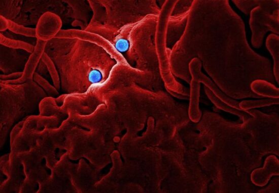
Extracellular Vesicle Communication Analysis: Tiny particles released by cells transport biological information between tissues
Anshika Jain - November 19, 2026The human body contains billions of cells that continuously exchange information to coordinate biological activities. Cells communicate through chemical signals, hormones, electrical impulses and direct physical interactions.…

Glycoprotein Structure Prediction: Changes in Protein-linked sugar structures
Anshika Jain - November 19, 2026Proteins are responsible for many essential biological activities, including cellular communication, immune defence, molecular transport, tissue development and the regulation of biochemical reactions. However, proteins do not…

Cellular Autophagy Regulation Intelligence: Cells recycle damaged components
Anshika Jain - November 19, 2026Every living cell continuously performs a complex maintenance process to preserve its internal environment, remove damaged structures and maintain normal biological activity. Proteins become misfolded, cellular membranes…
Most from this category


Behavioural Adaptation Under Uncertainty: People maintain health routines
Anshika Jain - November 19, 2026
Wellness Decision Overload Architecture: Conflicting health information changes
Anshika Jain - November 18, 2026
Health Behaviour Switching Costs: Mental effort replace unhealthy routines
Anshika Jain - November 18, 2026
RNA Modification Mapping: Chemical Modifications to RNA Influence
Anshika Jain - November 19, 2026Behavioural Adaptation Under Uncertainty: People maintain health routines
Anshika Jain - November 19, 2026Wellness Decision Overload Architecture: Conflicting health information changes
Anshika Jain - November 18, 2026


RNA Modification Mapping: Chemical Modifications to RNA Influence
November 19, 2026
.png)
Leave a Comment
You must be logged in to post a comment.