
3D Bioprinting: The Future of Manufacturing Human Tissues?

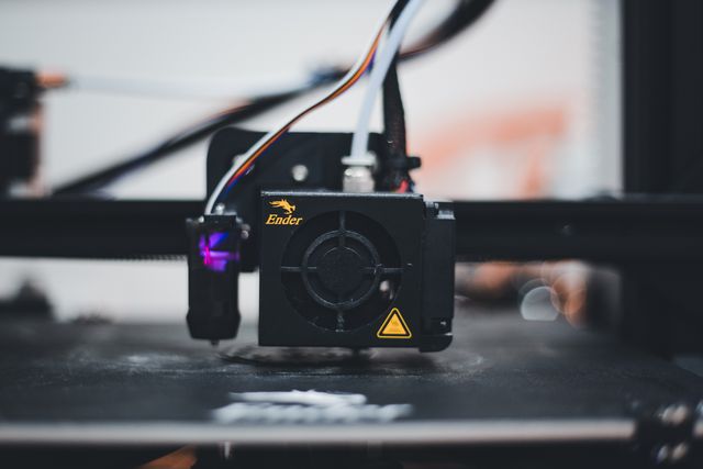
Three-dimensional bioprinting has emerged as one of the technologies attempting to address this challenge. Unlike conventional 3D printing, which typically deposits plastics, metals or other non-living materials, 3D bioprinting involves the controlled placement of living cells, biomaterials and biological components to construct tissue-like structures. The objective is not simply to produce a physical shape but to create an environment in which cells can survive, organize, mature and eventually perform tissue-specific functions.
The technology combines principles from tissue engineering, stem-cell biology, materials science, computer-aided design, robotics and regenerative medicine. Researchers can use digital models to determine where different cells and biomaterials should be placed, creating structures with spatial organization that would be difficult to achieve using conventional tissue-engineering methods. Current research includes bioprinted models of skin, cartilage, bone, vascular structures, cardiac tissue, liver tissue and other biological systems, while the longer-term ambition is to create increasingly complex tissues and potentially transplantable organs.
However, the phrase “printing human organs” can create unrealistic expectations. Scientists are not yet routinely producing fully functional, transplantable human hearts, kidneys or livers on demand. Major challenges involving vascularization, maturation, mechanical strength, long-term survival, immune compatibility, manufacturing consistency and regulatory approval remain. The significance of 3D bioprinting therefore lies not only in its distant possibility of manufacturing replacement organs but also in its growing ability to create useful human tissue models for research and regenerative medicine.
What Is 3D Bioprinting?
3D bioprinting is an additive manufacturing approach in which biological materials are deposited in controlled patterns to create three-dimensional structures. The process generally begins with a digital representation of the desired tissue architecture. Researchers then determine the appropriate cells, biomaterials and printing method required to reproduce aspects of that structure.
The material used in bioprinting is often called a bioink. Unlike ordinary printer ink, a bioink may contain living cells suspended within a supportive biomaterial. Depending on the intended tissue, the formulation can be designed to provide appropriate mechanical properties, support cell survival and allow cells to attach, migrate, differentiate or produce their own extracellular matrix.
The printing process can involve extrusion-based systems, droplet-based approaches, laser-assisted techniques or light-based methods such as projection-based bioprinting. Each approach provides different combinations of printing speed, resolution, material compatibility and cell-handling capabilities. Recent research has expanded the range of techniques available for depositing cells and biological building blocks with increasingly precise spatial control.
The ultimate objective is therefore more sophisticated than simply printing a biological shape. A successful bioprinted tissue needs to possess an appropriate architecture and provide a biological environment in which living cells can survive and develop into functional tissue.
Why Manufacturing Human Tissue Is So Difficult
The human body is an extraordinarily complex biological system. A natural organ is not simply a collection of cells arranged in a particular shape. It contains multiple cell types, extracellular matrix, blood vessels, nerves, immune components and biochemical signals that interact continuously.
A bioprinted tissue must therefore reproduce several levels of organization simultaneously. Cells must be positioned appropriately, but they must also communicate with neighbouring cells. The supporting material must provide structural stability without interfering with biological processes. Nutrients and oxygen must reach cells, waste products must be removed, and the tissue must be capable of integrating with the surrounding body after implantation.
This is one reason why creating a replacement organ is substantially more complicated than producing a conventional 3D-printed medical device. A printed implant can sometimes provide mechanical support without being alive. A bioprinted organ must remain biologically active and function within a dynamic physiological environment.
Researchers are therefore increasingly treating bioprinting as a complete biofabrication process rather than simply a printing technique. The printer, bioink, cell source, maturation environment and post-printing biological processes all influence the final result.
The Importance of Bioinks
Bioinks are at the centre of 3D bioprinting. A suitable bioink must satisfy several competing requirements. It needs to be printable, meaning it should flow through the selected printing system in a controlled manner. At the same time, it needs to provide a supportive environment for living cells.
The material must also achieve an appropriate balance between strength and biological compatibility. A material that is mechanically strong may not necessarily provide an environment that cells can tolerate. Conversely, a very soft material may support cells but fail to provide sufficient structural integrity.
Researchers are investigating natural materials, synthetic polymers and combinations of different materials to create increasingly sophisticated bioinks. Some approaches use components inspired by the extracellular matrix, while others incorporate growth factors or other biological signals that influence cell behaviour.
The development of better bioinks is particularly important because different tissues have very different requirements. Bone needs substantial mechanical strength, while soft tissues such as brain tissue require very different physical characteristics. A universal bioink is therefore unlikely to be sufficient for every application.
Extrusion, Droplet and Light-Based Bioprinting
Different bioprinting technologies offer different advantages. Extrusion-based bioprinting is one of the most widely studied approaches. In this method, bioink is deposited through a nozzle, allowing researchers to build structures layer by layer. It can handle relatively high cell densities and a range of biomaterials, although increasing printing speed or resolution can introduce biological and engineering trade-offs.
Droplet-based bioprinting instead deposits controlled droplets of biological material. This approach can provide high-throughput manipulation of biological components and can be useful when researchers need precise control over small volumes. Recent methodological research has emphasized its ability to manipulate cells and tissue building blocks at relatively fine scales.
Light-based systems use projected patterns or other optical methods to selectively solidify photosensitive materials. These technologies can potentially achieve high printing speeds and fine spatial control. However, the interaction between light, biomaterials and living cells introduces its own technical challenges, including limitations in resolution and the need to maintain cell viability during fabrication.
The continued development of these approaches means that bioprinting is becoming a family of technologies rather than a single standardized process.
From Cells to Structured Tissue
One of the most important advantages of bioprinting is spatial control. Researchers can theoretically determine where different cell types and materials should be positioned within a construct.
This capability becomes important when recreating tissues containing multiple cellular populations. For example, a tissue may require different types of cells to occupy different regions while remaining connected through extracellular structures and vascular networks.
Organoids and other self-organizing biological structures can also be incorporated into bioprinting strategies. Rather than attempting to manually print every individual cell, researchers can combine the precision of additive manufacturing with the natural ability of cells to self-organize. This hybrid approach is attracting attention because complex human tissues require both engineering control and biological self-assembly.
This represents an important shift in the field. The future of bioprinting may not involve a printer independently constructing every detail of a tissue. Instead, engineering systems may provide the initial architecture while biological processes complete the organization and maturation.
Bioprinting Skin, Cartilage and Bone
Some of the most accessible applications of bioprinting involve tissues that are structurally less complex than major internal organs. Skin, cartilage and bone have therefore received considerable research attention.
Bioprinted skin could potentially support wound healing and reconstruction by creating tissue structures containing appropriate cells and biomaterials. Researchers are also investigating bioprinted cartilage for applications involving damaged joints and reconstructive surgery.
Bone presents different requirements because it must withstand mechanical forces. Bioprinting can potentially create customized structures that combine appropriate geometry with biological components that support tissue regeneration.
These applications illustrate why the future of bioprinting does not necessarily depend on immediately producing entire organs. Creating smaller, specialized tissue replacements could provide important clinical applications while researchers continue addressing the much greater complexity of whole-organ fabrication. Reviews of current research identify skin, musculoskeletal tissues, cartilage and bone among important areas of ongoing development.
The Challenge of Vascularization
Vascularization is one of the biggest obstacles to manufacturing thick, living tissues. Cells require oxygen and nutrients, and they must release metabolic waste. In the human body, dense networks of blood vessels solve this problem by bringing circulation deep into tissues.
A large bioprinted structure cannot simply depend on nutrients diffusing from its outer surface. Cells located deep inside a thick construct can become deprived of oxygen and nutrients if adequate perfusion is not established.
Researchers are therefore working on methods for creating interconnected vascular structures inside bioprinted tissues. These approaches include printing channels, incorporating vascular cells and developing materials that encourage blood-vessel formation.
The challenge is particularly important for whole-organ bioprinting. A functional organ would require an extremely complex vascular network capable of connecting with the patient’s circulatory system. Current research continues to identify vascularization, tissue thickness and long-term perfusion as major barriers to creating clinically functional large tissues.
Can Bioprinted Tissues Integrate With the Body?
Creating tissue outside the body is only one part of the problem. After implantation, the printed structure must interact with the patient’s existing biological environment.
Cells need to survive and integrate with surrounding tissues. Blood vessels may need to connect with the patient’s circulation. Nerves may need to establish functional connections. The immune system must also tolerate the implanted material and cells.
Patient-derived cells could potentially reduce certain compatibility problems because the biological material originates from the individual receiving the treatment. However, using patient-derived cells introduces additional manufacturing complexity, including cell collection, expansion, quality control and individualized production.
Researchers are also studying biomaterials and tissue-engineering strategies designed to encourage appropriate integration while minimizing unwanted immune responses. These biological interactions remain central to the clinical translation of bioprinted tissues.
3D Bioprinting and Drug Development
The importance of 3D bioprinting extends beyond transplantation. Bioprinted tissues can also become experimental models for studying disease and testing drugs.
Traditional laboratory cell cultures often lack the three-dimensional architecture of human tissues. Bioprinted models can introduce spatial organization and multiple cell types, potentially providing researchers with more physiologically relevant experimental environments.
Cancer research is one area where this approach is particularly interesting. Researchers can construct tumour models containing different cell populations and extracellular environments, then investigate how they respond to potential treatments.
Bioprinted tissues could also contribute to toxicity testing. Instead of examining how a drug affects only isolated cells, researchers can study its effects within a structured tissue model. This could complement existing laboratory and animal research and provide additional information during drug development.
The field is therefore relevant even if fully transplantable organs remain a distant objective. The ability to manufacture standardized biological models could itself have significant implications for biomedical research.
Artificial Intelligence and the Future of Bioprinting
Artificial intelligence is increasingly being connected with bioprinting because the process involves large amounts of biological and engineering data.
AI systems could help researchers optimize bioink formulations, printing parameters, cell distributions and tissue architectures. Computer vision could monitor printed structures and identify changes in shape, cell viability or tissue development. Machine-learning models could also help identify relationships between printing conditions and biological outcomes.
The combination of AI and bioprinting could eventually produce more adaptive manufacturing systems. Instead of following a fixed printing protocol, future systems might continuously analyse the developing tissue and adjust parameters accordingly.
Recent research has specifically identified intelligent process control and machine learning as emerging directions for improving bioprinting and accelerating its translation toward clinically relevant tissue fabrication.
This could transform bioprinting from a relatively manual laboratory process into a more automated and data-driven form of biological manufacturing.
Why Printing a Whole Human Organ Is Still Difficult
The idea of printing a complete human heart, liver or kidney is scientifically compelling, but it represents a far greater challenge than printing a simpler tissue structure.
A functional organ must contain many specialized cell types arranged in precise anatomical relationships. It must have an extensive vascular network, appropriate mechanical properties, electrical or biochemical functionality where necessary, and the ability to respond dynamically to the body’s signals.
The organ must also survive for years rather than simply functioning temporarily in a laboratory environment. Long-term stability, maturation and integration remain major unresolved questions.
Current reviews emphasize that clinically relevant bioprinted tissues still need to demonstrate sufficient functionality, longevity, biomechanical performance, vascularization and, in some applications, innervation. The field is therefore progressing toward increasingly sophisticated tissue constructs, but routine manufacturing of complete transplantable human organs remains a research objective rather than an established clinical capability.
Regulation, Safety and Manufacturing Standards
Another major challenge is regulation. A conventional medical device may be manufactured using standardized materials and processes, but a bioprinted tissue can contain living cells, biomaterials and biological signals whose properties may change over time.
Regulators therefore need to consider questions involving cell identity, viability, sterility, biomaterial safety, manufacturing consistency, immune reactions and long-term clinical outcomes. The individualized nature of some bioprinted products also creates challenges for quality control and scalable manufacturing.
Researchers have highlighted the need for clearer standards and regulatory frameworks capable of addressing the unique characteristics of living bioprinted products. Standardization is especially important because differences in printers, bioinks, cell sources and fabrication protocols can produce substantially different biological outcomes.
The development of robust manufacturing standards will be essential if bioprinting is eventually to move from specialized research laboratories into routine clinical environments.
Ethical and Economic Considerations
Scientific feasibility is not the only factor that will determine the future of 3D bioprinting. Ethical and economic questions will also become increasingly important.
Patient-derived cells raise questions about consent, biological ownership, privacy and the future use of stored cellular material. If a patient’s cells are used to manufacture a personalized tissue, researchers and healthcare providers will need clear systems for managing biological data and manufacturing records.
Cost is another consideration. Advanced bioprinting equipment, specialized bioinks, controlled laboratory environments, skilled personnel and quality-control systems can make production expensive. A personalized bioprinted tissue may be scientifically possible but still difficult to manufacture affordably and consistently at large scale.
Clinical adoption will therefore depend not only on whether a bioprinted tissue works, but also on whether it can be produced safely, reliably and economically.
Could Bioprinting Eventually Reduce the Organ Shortage?
The shortage of transplantable organs is one of the reasons bioprinting has attracted so much attention. In theory, manufacturing tissues from a patient’s own cells could provide an alternative to waiting for a compatible donor.
However, this possibility should be understood as a long-term research direction rather than an immediate solution. Producing a structurally accurate organ is only the beginning. The organ must mature, establish vascular and functional connections, integrate with the recipient and remain healthy for a long period.
Researchers are therefore pursuing multiple strategies simultaneously. Some focus on complete organs, while others work on smaller tissue patches, vascular structures, organ models and regenerative scaffolds. This gradual approach could allow individual technological advances to become clinically useful before the much larger challenge of complete organ fabrication is solved.
The Future of Human Tissue Manufacturing
The future of 3D bioprinting will likely involve the convergence of several technologies rather than improvements in printing alone. Stem-cell biology can provide increasingly capable cellular building blocks. Biomaterials research can produce better bioinks. Organoid science can provide self-organizing tissue structures. Artificial intelligence can optimize manufacturing and analyse biological responses. Microfluidic systems can reproduce controlled physiological environments, while advanced imaging can guide increasingly precise fabrication.
This convergence could lead to a new form of biological manufacturing in which tissue is designed digitally, constructed using living materials and then matured under controlled conditions.
Researchers are also exploring increasingly sophisticated strategies for multi-material and multi-modal bioprinting. Such systems could eventually allow different tissue regions to be produced using different materials and cell populations within a single construct. The long-term objective is to reproduce the hierarchical organization found in natural tissues with increasing precision.
Conclusion
3D bioprinting represents a significant intersection between engineering and biology. By enabling researchers to position living cells and biomaterials in controlled three-dimensional structures, the technology provides new possibilities for tissue engineering, regenerative medicine, disease modelling and drug research.
The idea of manufacturing replacement human organs is one of the most ambitious possibilities associated with the technology, but the scientific reality remains more complex. Vascularization, cellular maturation, mechanical strength, long-term function, immune compatibility, reproducibility, manufacturing scale and regulatory approval continue to limit clinical translation. Current research therefore focuses heavily on creating functional tissue components and improving the reliability of bioprinted constructs rather than simply attempting to print complete organs.
The most important development may ultimately be the gradual transformation of how scientists manufacture and study human tissue. Bioprinting could make it possible to create increasingly realistic tissue models for research, customized structures for regenerative medicine and, eventually, more complex biological replacements.
Whether it will ultimately become a routine method for manufacturing entire human organs remains an open scientific question. What is increasingly clear is that 3D bioprinting has moved beyond the idea of simply printing shapes. It is becoming a sophisticated biofabrication technology in which cells, biomaterials, digital design, engineering and biological self-organization work together. As these fields continue to converge, the ability to manufacture living human tissue may become an increasingly important part of the future of medicine.
Online Internship with Certificate
You may be interested
Stem Cell Engineering and the Search for New Regenerative Therapies
Anshika Jain - October 1, 2026The human body possesses a remarkable capacity for repair. Skin can close wounds, bones can remodel after injury, blood cells are continuously replaced, and tissues throughout the…
Organoids: How Miniature Human Organs Are Changing Medical Research
Anshika Jain - October 1, 2026Medical research has traditionally depended on a combination of laboratory cell cultures, animal models, clinical observations, and human studies. Each approach has contributed enormously to scientific progress,…
Liquid Biopsy Technology: Can Blood Samples Reveal Molecular Signals From Tumors?
Anshika Jain - October 1, 2026Cancer diagnosis and monitoring have traditionally depended heavily on tissue biopsies, imaging, and laboratory testing. A tissue biopsy can provide valuable information because it allows pathologists to…
Most from this category

Personalized Oncology: How Tumor Genetics Is Changing Cancer Treatment Strategies
Anshika Jain - October 1, 2026
Multi-Omics Medicine: Combining Genomics, Proteomics, Metabolomics and Other Biological Layers
Anshika Jain - October 1, 2026
Immunometabolism: How Cellular Energy Systems Influence Immune Function
Anshika Jain - September 30, 2026
The Gut Barrier: Why Intestinal Permeability Has Become a Major Area of Biomedical Research
Anshika Jain - September 30, 2026
Personalized Oncology: How Tumor Genetics Is Changing Cancer Treatment Strategies
Anshika Jain - October 1, 2026Multi-Omics Medicine: Combining Genomics, Proteomics, Metabolomics and Other Biological Layers
Anshika Jain - October 1, 2026Immunometabolism: How Cellular Energy Systems Influence Immune Function
Anshika Jain - September 30, 2026



.png)
Leave a Comment
You must be logged in to post a comment.