
Cellular Stress Communication Networks: Stressed Cells Influence Neighbouring Healthy Cells

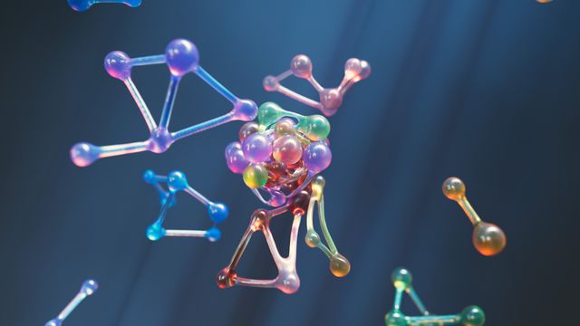
Cells do not respond to stress in isolation. When a cell experiences DNA damage, oxidative stress, inflammation, nutrient deprivation or exposure to harmful substances, it can release molecular signals that influence the behaviour of nearby cells. These signals may encourage neighbouring cells to activate protective mechanisms, alter their metabolism, enter a temporary growth-arrest state or, in some circumstances, develop damaging responses of their own. This communication can help tissues coordinate repair, but it can also spread inflammation and contribute to disease.
The emerging concept of Cellular Stress Communication Networks focuses on understanding how these signals travel between cells, how neighbouring cells interpret them and how local stress can influence the behaviour of an entire tissue. Artificial intelligence could make this complex process easier to investigate by analysing molecular interactions, spatial patterns, gene activity and changes in cell behaviour over time. By combining these sources of information, AI may help researchers identify how stress signals originate, which cells receive them and whether communication supports tissue recovery or contributes to long-term damage.
A cellular stress communication network can be understood as a system of connected sender cells, signalling molecules and receiver cells. A stressed cell acts as a sender by releasing or displaying biological signals. Nearby cells act as receivers when they detect these signals through specific receptors or other communication mechanisms. The receiving cells then activate internal pathways that influence their behaviour. These responses can create additional signals, allowing the original message to spread through a tissue.
This process is not necessarily harmful. Communication between stressed and healthy cells is an essential part of tissue maintenance, immune defence and wound healing. The biological outcome depends on the type of stress, the identity of the cells, the signals released, the duration of exposure and the condition of the surrounding tissue.
How Stressed Cells Communicate With Healthy Neighbours
Cells use several mechanisms to exchange information. One of the most important is chemical signalling through molecules such as cytokines, chemokines, growth factors and other secreted proteins. A stressed cell can release these substances into its surrounding environment. They travel through the extracellular space and interact with receptors on neighbouring cells, activating specific intracellular signalling pathways.
Cytokines are particularly important in stress-related communication. Molecules such as interleukin-6 and interleukin-8 can influence inflammatory responses, immune-cell recruitment and tissue behaviour. Depending on the biological context, their release may help coordinate repair or contribute to persistent inflammation. Growth factors and transforming growth factor beta can also influence cell survival, proliferation, differentiation and tissue remodelling.
Another important communication mechanism involves extracellular vesicles. These are small membrane-bound structures released by cells that can carry proteins, lipids, messenger RNA, microRNA and other molecular material. When neighbouring cells take up these vesicles, their internal signalling and gene-expression patterns may change. Extracellular vesicles therefore provide a way for stressed cells to influence other cells without requiring direct physical contact.
Direct cell-to-cell communication can also occur through gap junctions. These specialised connections allow certain small molecules and ions to pass between neighbouring cells. They are particularly important in tissues where coordinated activity is necessary, such as cardiac and some epithelial tissues. Stress-related changes in gap-junction communication may influence how cells coordinate their responses.
Cells can also release damage-associated molecular patterns, commonly known as DAMPs. These molecules or cellular components become available outside cells following damage or disruption. They can activate receptors involved in innate immune responses and alert surrounding tissues to potential danger. The resulting response may promote defence and repair, although excessive or prolonged activation can contribute to tissue injury.
These mechanisms can operate simultaneously. A stressed cell may release cytokines, extracellular vesicles and other signalling molecules while changing its direct interactions with neighbouring cells. Understanding their combined effects is one of the major challenges that AI-assisted research could help address.
The Role of Cellular Senescence in Stress Signal Propagation
Cellular senescence is one of the most studied examples of how a stressed cell can influence its surrounding environment. Senescence is a cellular state in which a cell undergoes a relatively stable arrest of cell division, often following DNA damage, oncogene activation or other forms of stress. This response can prevent damaged cells from continuing to divide, thereby helping to limit the development of certain cancers.
However, senescent cells are not biologically inactive. Many develop a characteristic pattern of secreted molecules known as the senescence-associated secretory phenotype, or SASP. This can include inflammatory cytokines, chemokines, growth factors and enzymes that modify the surrounding extracellular environment.
The SASP can have beneficial effects in specific situations. It may recruit immune cells to remove damaged cells, support tissue remodelling and contribute to wound healing. Yet persistent SASP activity can also influence neighbouring healthy cells in less favourable ways. It may promote inflammation, alter tissue structure, encourage abnormal cellular behaviour and contribute to the spread of senescence-like characteristics under certain experimental conditions.
Research increasingly recognises that senescent cells can influence biological processes beyond their immediate surroundings. A 2025 review in Physiology examines how senescence-associated secretory factors, metabolites, extracellular vesicles, immune cells and other communication routes can influence interorgan signalling and systemic health.
A 2026 review on intercompartmental communication in senescence further explores how changes in intracellular transport, vesicle trafficking and communication between organelles can contribute to oxidative stress, impaired cellular maintenance and SASP activation.
For AI researchers, senescence provides a useful model for studying stress communication because it involves identifiable changes in gene expression, molecular secretion and neighbouring-cell behaviour. A computational system could compare the molecular profiles of senescent cells with those of nearby non-senescent cells and investigate whether specific signalling patterns are associated with changes in the receiving population.
Why Understanding Neighbouring Healthy Cells Is Difficult
One of the central challenges in cellular stress research is distinguishing between the original stressed cell and the cells responding to its signals. A tissue contains many cell types, and each may react differently to the same molecular message. A cytokine released by a damaged epithelial cell, for example, may activate an immune cell differently from how it affects a neighbouring epithelial cell or a fibroblast.
The same signal can also produce different outcomes depending on the condition of the receiving cell. A healthy cell may activate a protective response, while a cell already experiencing metabolic or oxidative stress may develop a stronger inflammatory response. The receptor levels, internal signalling pathways, genetic background and surrounding chemical environment all influence the final outcome.
Distance is another important factor. Some signals act over very short distances, while others can travel further through tissue fluids or circulation. A cell located immediately beside a stressed cell may experience a different concentration of signalling molecules from a cell several tissue layers away. Tissue structure, blood flow, extracellular matrix and the presence of immune cells can alter how signals move.
Time adds another layer of complexity. A short period of stress may trigger a temporary protective response, whereas repeated or prolonged exposure can produce persistent changes. A single tissue sample provides only a snapshot of this process. Understanding how stress communication develops requires measurements across multiple time points.
These challenges explain why examining isolated cells is not always sufficient. Researchers need to understand both the molecular characteristics of individual cells and their relationships within the surrounding tissue.
How AI Can Decode Cellular Stress Communication Networks
Artificial intelligence can help analyse cellular communication by integrating information that is too complex to interpret through individual measurements. Machine learning models can examine gene-expression patterns, molecular signals, cell identities and spatial relationships to identify possible connections between stressed cells and their neighbours.
One important source of information is single-cell RNA sequencing. This technology measures gene-expression patterns at the level of individual cells, helping researchers identify cell populations with different biological states. By comparing stressed and unstressed cells, researchers can identify changes in the expression of genes associated with cytokines, receptors, inflammatory pathways and cellular repair.
AI models can use these patterns to predict possible communication between sender and receiver cells. For example, a stressed cell population may show increased expression of a signalling molecule, while a neighbouring population expresses the corresponding receptor. Computational tools can use curated ligand–receptor databases to identify plausible interactions and estimate their potential relevance.
However, detecting a ligand and its receptor does not prove that communication actually occurred. The proteins may not be produced, secreted, available or active under the conditions being studied. AI-generated interactions should therefore be treated as hypotheses that require experimental validation.
Spatial transcriptomics provides an additional advantage because it connects gene-expression measurements with the physical location of cells inside tissue. Unlike conventional single-cell sequencing, which generally requires tissue dissociation, spatial methods preserve information about where cells are positioned relative to one another. This allows researchers to investigate whether a potential sender and receiver are located close enough for a particular interaction to be biologically plausible.
A 2026 review in Briefings in Bioinformatics describes two major computational approaches to spatial cell–cell communication analysis. One identifies spatial patterns, including the co-location of signalling molecules and receptor-expressing cells. The other examines how the local cellular environment may influence the gene-expression state of receiving cells. The review also highlights the need for better validation, benchmarking and integration of multiple biological data types.
Using Graph Neural Networks to Model Cellular Relationships
Cellular communication is naturally suited to network-based analysis. In a biological network, individual cells can be represented as nodes, while their potential communication relationships are represented as connections or edges. Each node can contain information about cell type, gene expression, stress status, receptor availability and spatial position.
Graph neural networks could help analyse these relationships. These AI models learn from both the characteristics of individual nodes and the connections between them. In a tissue model, they could examine how the state of a stressed cell relates to the molecular characteristics of neighbouring cells.
For example, researchers could construct a network containing damaged epithelial cells, nearby fibroblasts, immune cells and healthy epithelial cells. The model could examine which cell populations express particular signalling molecules, which neighbouring populations possess corresponding receptors and how their gene-expression profiles change across different conditions.
A more advanced model could analyse higher-order relationships. Instead of studying only one stressed cell and one neighbouring cell, it could examine how a group of stressed cells influences a local tissue environment. It might identify whether immune-cell recruitment, fibroblast activation and epithelial responses occur together as part of a broader communication pattern.
This approach could be useful in cancer research, chronic inflammation and tissue repair. However, network connections inferred from gene expression and spatial proximity do not automatically establish direct biological causation. Graph-based predictions need to be tested through laboratory experiments that manipulate the proposed signalling pathways.
Integrating Multi-Omics Data for a More Complete Picture
Gene expression alone cannot explain every aspect of cellular communication. A cell may express a signalling gene without releasing a functional protein. A receptor may be present but inactive. A molecule may be secreted but rapidly degraded or blocked by its surrounding environment. Understanding these processes requires information from multiple levels of cellular biology.
Multi-omics integration could help address this limitation. Transcriptomics provides information about RNA expression, proteomics measures proteins, metabolomics examines small molecules and lipidomics studies lipid profiles. Epigenomic measurements can reveal changes in gene regulation, while imaging can show where cells and molecular structures are located.
AI could combine these datasets to develop a more complete representation of stress communication. For example, transcriptomic data might identify increased expression of an inflammatory signalling molecule, while proteomic analysis could determine whether its protein is present. Spatial imaging could establish whether the producing cells are close to potential receiver cells, and metabolomic measurements could reveal whether the surrounding tissue has developed a changed biochemical environment.
The integration of these data types may also help researchers identify communication pathways that are not captured by conventional ligand–receptor analysis. Cells exchange information through extracellular vesicles, metabolic products, mechanical interactions and other mechanisms. A multi-omics framework could help explore how these different processes interact, although each proposed mechanism would require appropriate experimental evidence.
Potential Applications in Cancer, Inflammation and Tissue Repair
Cancer research is an important application area because tumour cells do not develop independently of their surroundings. Tumours contain immune cells, fibroblasts, blood vessels and other components that form a complex microenvironment. Stress within tumour cells can change the signals they release, influencing how neighbouring cells behave. These interactions may affect immune activity, tissue remodelling, treatment response and tumour progression.
AI-based communication mapping could help researchers identify signalling relationships between stressed tumour cells and surrounding immune or stromal cells. It may reveal spatial regions where particular interactions are more active and suggest which pathways should be investigated experimentally. Such analysis could eventually support the identification of new therapeutic targets, although computational predictions alone cannot establish treatment effectiveness.
Chronic inflammation is another relevant area. Repeated cellular stress can sustain the release of inflammatory mediators, changing the behaviour of nearby cells and potentially maintaining a cycle of tissue irritation. AI could compare tissue samples collected at different disease stages to investigate how signalling networks change as inflammation develops or resolves.
Tissue repair offers a contrasting application. Following an injury, damaged cells release signals that help recruit immune cells and coordinate repair processes. Fibroblasts, epithelial cells, endothelial cells and immune cells must communicate to restore tissue organisation. An AI model could examine how these interactions evolve and identify patterns associated with successful healing or excessive scar formation.
From Static Cellular Maps to Dynamic Communication Models
Many current cell–cell communication analyses rely on data collected at a single time point. Such studies can identify possible interactions, but they may not reveal how communication develops or changes over time. Cellular stress, however, is a dynamic process. The signals released immediately after injury may differ from those produced several hours or days later.
Future AI systems could combine time-series measurements with spatial information to reconstruct how communication networks change. Researchers might collect tissue measurements at several stages after a controlled stress exposure, then analyse changes in sender-cell activity, signalling molecule levels and receiver-cell responses.
Machine learning models could help identify which signals appear first, which cell populations respond next and whether those responses generate additional communication pathways. This could provide a more detailed understanding of how a local stress event develops into a wider tissue response.
Such models may also help distinguish temporary protective communication from persistent signalling associated with chronic inflammation or tissue damage. The distinction is important because the same molecule can have different effects depending on its concentration, duration of exposure and biological context.
The longer-term objective would be to move from static maps of possible cellular interactions towards dynamic models that represent the changing relationships between cells. Recent reviews of spatial transcriptomics emphasise that the field is moving towards more integrated, multilayered and dynamic approaches, while recognising that many existing methods still require stronger validation.
Scientific Limitations and the Need for Experimental Validation
Despite its potential, AI-based cellular stress communication analysis faces substantial limitations. The first is the quality and completeness of biological datasets. Single-cell sequencing may capture only a portion of the molecules present in a cell, while spatial transcriptomics technologies differ in resolution and sensitivity. Some methods identify expression across groups of cells rather than resolving every individual cell, which can complicate the interpretation of local interactions.
Another limitation is the incompleteness of ligand–receptor databases. Not every signalling interaction has been discovered or experimentally characterised. Some molecular relationships are highly dependent on tissue type, developmental stage or disease condition. A model trained on incomplete interaction databases may overlook important pathways or predict relationships that are not active in a particular biological environment.
AI models may also confuse correlation with causation. If two cell populations show coordinated gene-expression changes, the relationship could be caused by a shared environmental condition rather than direct communication between them. Spatial proximity and matching ligand–receptor expression strengthen a hypothesis but do not prove that a signal was transmitted or that it caused the observed response.
Experimental validation is therefore essential. Researchers may need to use cell-culture systems, organoids, co-culture experiments, molecular imaging and targeted genetic or pharmacological interventions to test predicted interactions. If blocking a proposed signalling pathway changes the behaviour of receiver cells in the expected way, this provides stronger evidence that the communication relationship is biologically meaningful.
Reproducibility is another concern. Results should be tested across independent samples, different experimental systems and, where possible, multiple computational methods. A model that performs well on one tissue dataset may not generalise to another because cell composition, tissue structure and signalling conditions differ.
The Future of AI-Driven Cellular Stress Intelligence
Cellular Stress Communication Networks offer a way to study disease and tissue health beyond the behaviour of individual cells. They focus on how stress signals move through biological environments and how groups of cells coordinate their responses. AI could make this investigation more powerful by combining molecular, spatial and temporal information into integrated communication models.
Future systems may help researchers identify which cells initiate stress responses, which neighbouring populations are most affected and which signalling pathways are associated with tissue protection or damage. They could also help prioritise molecular targets for experimental investigation and reveal why similar stress conditions produce different outcomes in different tissues.
The most important development will be the integration of computational predictions with biological evidence. More accurate spatial measurements, improved multi-omics technologies and stronger validation frameworks will be essential.
Ultimately, decoding cellular stress communication could improve scientific understanding of cancer, ageing, inflammation and tissue regeneration. Rather than viewing a damaged cell as an isolated unit, researchers may increasingly examine it as part of a wider network in which its signals influence the fate of neighbouring cells. AI could help reveal the structure and behaviour of these networks, bringing researchers closer to understanding how local cellular stress becomes a tissue-level response.
Online Internship with Certificate
You may be interested

Personal Biological Variability Fingerprinting: Distinguish Health Changes From Physiological Fluctuations
Anshika Jain - November 12, 2026The human body is constantly changing. Heart rate rises during physical activity, body temperature shifts throughout the day, hormone levels respond to sleep and stress, and blood…

Tissue-Level Communication Failures: AI Identify Hidden Disruptions in the Signals
Anshika Jain - November 12, 2026The human body functions as an interconnected biological network in which individual tissues continuously exchange information. The skin, muscles, brain, liver, heart, kidneys, immune system and other…

Subclinical Health Transition Detection: AI Recognise Disease Development
Anshika Jain - November 12, 2026Disease does not always begin when a person first experiences noticeable symptoms. In many conditions, biological changes develop gradually before an individual feels unwell or receives a…
Most from this category

Human Physiology Under Resource Competition: Body’s Prioritisation of Energy
Anshika Jain - November 12, 2026
Biological Redundancy Analysis: Which Body Systems Compensate When Another System Begins to Fail
Anshika Jain - November 12, 2026
Gut Virome Intelligence: Infect Bacteria Influence the Human Gut Ecosystem
Anshika Jain - November 6, 2026
Human–Environment Health Interaction Maps: AI Connect Indoor Lighting
Anshika Jain - November 6, 2026
Human Physiology Under Resource Competition: Body’s Prioritisation of Energy
Anshika Jain - November 12, 2026Biological Redundancy Analysis: Which Body Systems Compensate When Another System Begins to Fail
Anshika Jain - November 12, 2026Gut Virome Intelligence: Infect Bacteria Influence the Human Gut Ecosystem
Anshika Jain - November 6, 2026




.png)
Leave a Comment
You must be logged in to post a comment.