
Lab-Grown Tissues: How Biotechnology Is Redefining the Boundaries of Medical Research

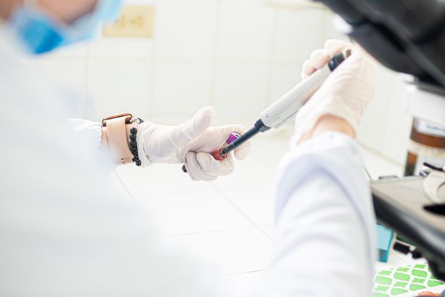
Modern medicine has made extraordinary progress in diagnosing and treating disease, but one fundamental problem remains difficult: how to replace or repair human tissue that has been severely damaged. Conventional treatments can manage symptoms, remove diseased tissue, or use artificial devices to compensate for lost biological functions. However, restoring living tissue requires more than mechanical replacement. It requires cells that can grow, communicate, organize themselves, respond to biological signals, and integrate with the surrounding body.
This challenge has created an increasingly important area of biotechnology focused on lab-grown tissues. Researchers are developing methods to grow human cells outside the body and organize them into structures that reproduce selected characteristics of natural tissues. These approaches combine stem cell biology, tissue engineering, biomaterials, organoid research, gene editing, bioprinting, microfluidics, and computational technologies.
Lab-grown tissues are not simply miniature versions of body parts. They are experimental biological systems that allow scientists to recreate aspects of human physiology in controlled environments. They can be used to investigate disease, test medicines, study development, and explore potential regenerative therapies.
The field is therefore redefining medical research by creating an intermediate space between traditional cell cultures and living human patients. Instead of studying disease only through isolated cells or relying exclusively on animal models, researchers can increasingly examine human-derived tissues in three-dimensional environments that more closely resemble biological conditions.
What Are Lab-Grown Tissues?
Lab-grown tissues are collections of living cells cultivated and organized outside the human body to reproduce some of the structural or functional characteristics of natural tissue. Depending on their purpose, they can range from relatively simple cellular structures to highly sophisticated three-dimensional models.
Some are generated from stem cells that are guided toward particular cell types. Others are derived from tissue samples collected from patients. In many cases, researchers combine several cell populations to recreate interactions that occur naturally inside an organ.
The term covers a broad spectrum of technologies. Organoids can reproduce selected characteristics of organs, engineered tissue scaffolds can support the development of specific tissue structures, and bioprinting can arrange cells and biomaterials into predefined patterns.
The common objective is to create biological models that are more representative of human tissue than conventional two-dimensional cell cultures.
Why Traditional Cell Cultures Are Not Always Enough
For decades, scientists have grown human and animal cells on flat laboratory surfaces to study biological processes. These cultures remain valuable because they are relatively inexpensive, controllable, and easy to manipulate.
However, a flat layer of cells does not reproduce the physical organization of most human tissues. In the body, cells exist within three-dimensional environments where they interact with neighboring cells, extracellular structures, blood vessels, immune cells, and biochemical gradients.
The spatial arrangement of cells can influence their behavior. Cells located at the center of a tissue may experience different oxygen and nutrient conditions from cells near the surface. Mechanical forces can affect cell differentiation, while signals from neighboring cells can alter gene expression and function.
Lab-grown three-dimensional tissues attempt to capture some of these complexities. This can make them particularly useful when researchers need to understand how cells behave collectively rather than individually.
Stem Cells as the Foundation of Tissue Engineering
Stem cells are central to many lab-grown tissue technologies because they can generate specialized cell populations. Pluripotent stem cells, including induced pluripotent stem cells, are particularly valuable because they can potentially be differentiated into many different cell types.
Researchers can expose stem cells to carefully controlled combinations of molecular signals that guide them toward specific developmental pathways. These processes attempt to reproduce some of the signaling events that occur naturally during human development.
Once specialized cells have been generated, researchers can encourage them to organize into three-dimensional structures. The resulting tissue models may reproduce selected features of the original biological tissue.
Patient-derived induced pluripotent stem cells provide another important possibility. A mature cell from an individual can be reprogrammed into a pluripotent state and then differentiated into another cell type for research. This can create disease models that carry aspects of the patient’s own genetic background.
Organoids: Self-Organizing Human Tissue Models
Organoids are among the most widely discussed examples of lab-grown tissue technology. They are three-dimensional structures created from stem cells or tissue-derived cells that reproduce selected characteristics of organs.
Scientists have developed organoid models representing aspects of tissues such as the intestine, brain, liver, kidney, lung, pancreas, and retina. These models do not reproduce complete human organs, but they can demonstrate patterns of cellular organization and biological activity that are difficult to achieve in conventional cell cultures.
One of the most interesting characteristics of organoids is self-organization. Under appropriate conditions, cells can arrange themselves into spatial structures without scientists manually positioning every individual cell.
This makes organoids valuable for developmental biology and disease research. Researchers can observe how tissues form, how genetic mutations alter development, and how cells respond to drugs or infectious agents.
Tissue Engineering: Giving Cells a Physical Environment
Cells require more than nutrients to develop into organized tissues. They interact continuously with the extracellular matrix, a complex network of proteins and molecules that provides structural support and biochemical signals.
Tissue engineering attempts to recreate aspects of this environment using scaffolds and biomaterials. A scaffold can provide physical structure while giving cells a surface on which to attach, migrate, and organize.
Researchers can design materials with different levels of stiffness, porosity, elasticity, and biochemical properties. These physical characteristics can influence cellular behavior.
Biomaterials may also be engineered to release signaling molecules at specific rates or locations. In this way, the scaffold becomes more than a passive support structure. It can become part of the biological instructions guiding tissue development.
3D Bioprinting and the Construction of Tissue Architecture
Three-dimensional bioprinting has introduced another approach to building lab-grown tissues. Instead of allowing cells to organize entirely on their own, bioprinting technologies can deposit cells, biomaterials, or combinations of both according to digitally designed patterns.
The concept resembles conventional 3D printing, but the materials are biological. Specialized bioinks can contain living cells combined with hydrogels or other supportive materials.
The attraction of bioprinting lies in its ability to create controlled spatial arrangements. Researchers can potentially place different cell types in specific locations and design structures that resemble aspects of natural tissue architecture.
However, printing a structure is not the same as creating functional living tissue. Printed cells must survive, communicate, mature, and establish appropriate biological networks after fabrication. Researchers therefore continue to investigate how printed tissues can develop the complex properties required for real biological function.
The Vascularization Problem
One of the biggest challenges facing lab-grown tissue research is vascularization. Human tissues require networks of blood vessels to deliver oxygen and nutrients and remove waste products.
Small cellular structures can sometimes survive through diffusion, but larger tissues quickly encounter limitations. Cells located far from a nutrient source may become deprived of oxygen and begin to deteriorate.
Researchers are exploring several strategies to overcome this problem. These include incorporating vascular cells, creating microfluidic channels, designing materials that support vessel formation, and developing engineered vascular networks.
Solving vascularization is essential if researchers hope to create increasingly large and functional tissues. It is also one of the major barriers separating laboratory tissue models from fully functional transplantable organs.
Lab-Grown Tissues in Drug Discovery
Drug development is another area where lab-grown tissues could have a significant impact. Pharmaceutical researchers traditionally rely on combinations of molecular assays, cell cultures, animal models, and clinical trials.
Although these approaches are essential, experimental results from simplified models do not always predict how a medicine will behave in humans. Lab-grown human tissues could provide an additional level of biological complexity before a candidate therapy reaches clinical testing.
Liver tissue models can be used to investigate potential drug toxicity. Intestinal models can help researchers study absorption and tissue responses. Cardiac models can be examined for changes in electrical or contractile behavior.
These models may allow researchers to identify ineffective or potentially harmful compounds earlier in the development process, potentially reducing the cost and time associated with late-stage failures.
Personalized Medicine Through Patient-Derived Tissues
Perhaps one of the most transformative possibilities is the development of patient-specific tissue models. Cells collected from an individual can potentially be expanded, reprogrammed, and used to create laboratory tissue that reflects aspects of that person’s biology.
In cancer research, tumor-derived organoids can be exposed to different drugs to study treatment responses. Researchers can compare how tissue models from different patients respond to the same therapy.
This approach could eventually contribute to more personalized treatment strategies. Instead of selecting therapies solely from population-level evidence, clinicians may one day have additional laboratory evidence showing how a patient’s own biological material responds to particular interventions.
However, this potential remains an area of active research. Laboratory responses do not necessarily reproduce everything that happens inside a human body, where immune, hormonal, vascular, and metabolic systems interact with tumors and other tissues.
Lab-Grown Tissues and Disease Modeling
Human tissue models can provide researchers with opportunities to study diseases in environments that more closely resemble human biology.
Genetic diseases can be modeled by creating tissues from cells carrying disease-associated mutations. Researchers can then observe how those mutations affect tissue development or function.
Infectious disease research can also benefit from tissue models. Scientists can expose lab-grown tissues to pathogens and observe how infection begins, spreads, and triggers cellular responses.
Neurological research is another important area. Brain-related organoids and neural tissue models can help researchers investigate developmental processes and certain disease mechanisms that cannot be easily observed directly in humans.
The ability to manipulate these models experimentally makes them particularly useful for testing hypotheses about disease progression.
Lab-Grown Tissues and Cancer Research
Cancer research has become one of the most active applications of three-dimensional tissue models. Tumors contain diverse populations of cells and interact with their surrounding environment. These characteristics can be difficult to reproduce in conventional cell cultures.
Patient-derived cancer organoids can retain some of the genetic and molecular features of the original tumor. Researchers can use these models to investigate tumor growth, treatment response, resistance, and biological differences between patients.
Cancer tissue models can also be combined with other technologies. Genetic sequencing can reveal mutations, while drug-screening systems can test therapeutic compounds. Artificial intelligence can analyze images and molecular datasets to identify patterns associated with treatment response.
Such integrated systems could contribute to more detailed models of tumor biology and accelerate the development of targeted therapies.
Organ-on-a-Chip Systems
Organ-on-a-chip technology is expanding the capabilities of lab-grown tissues by introducing controlled microenvironments that mimic aspects of human physiology.
These systems use miniature devices containing channels through which fluids can flow around living cells or tissue models. Researchers can control mechanical forces, nutrient delivery, oxygen levels, and chemical exposure.
A lung-on-a-chip, for example, can be designed to reproduce certain physical conditions associated with breathing and interactions between lung cells and circulating fluids. Other systems are being developed to model tissues such as the liver, intestine, kidney, and blood-brain barrier.
Combining organoids or engineered tissues with microfluidic systems could create more dynamic laboratory environments that reproduce interactions impossible to capture in static cultures.
Artificial Intelligence and Lab-Grown Tissue Research
The increasing complexity of lab-grown tissues has created a growing role for artificial intelligence. Advanced tissue models can produce large datasets through microscopy, genetic sequencing, molecular profiling, and functional measurements.
AI systems can analyze high-resolution images to identify changes in tissue structure, cell morphology, growth patterns, and responses to experimental treatments. Machine learning can also help researchers classify tissue states and identify subtle differences between healthy and diseased models.
Automation could further accelerate the field. Robotic systems can potentially control cell cultures, perform repeated experiments, capture images, and process experimental data.
The combination of biological engineering and AI could make tissue research more scalable while reducing some of the variability associated with manual laboratory procedures.
The Challenge of Reproducing Real Human Biology
Despite impressive advances, lab-grown tissues remain simplified representations of the human body. Even highly sophisticated models generally lack some combination of mature blood vessels, nerves, immune cells, endocrine interactions, mechanical forces, and systemic metabolism.
A tissue may behave realistically in isolation but respond differently when placed within the complex environment of a living organism.
This limitation is particularly important for drug testing. A compound may appear safe in an isolated tissue model but behave differently when metabolized by the liver or processed by other organs.
Researchers are therefore increasingly interested in connecting multiple tissue models. The concept of interconnected human tissue systems could eventually allow scientists to study how a drug or disease affects several biological systems simultaneously.
Ethical and Regulatory Considerations
The growth of lab-grown human tissues also raises important ethical questions. When tissues are created from patient-derived cells, issues surrounding informed consent, genetic information, ownership, and future use of biological materials must be addressed.
Brain-related tissue models raise additional questions as scientists explore increasingly sophisticated neural structures. Although current models do not reproduce the full complexity of a human brain, continued technological development makes ethical discussion increasingly important.
Regulatory frameworks must also evolve alongside the technology. Researchers need clear standards for manufacturing, quality control, safety testing, and clinical translation.
Scientific progress and responsible governance must therefore develop together as lab-grown tissues become more advanced.
The Future of Lab-Grown Human Tissues
The next stage of the field is likely to involve increasing integration between several technologies. Stem cells can provide the cellular foundation, gene editing can modify specific biological properties, biomaterials can provide structural environments, bioprinting can control spatial organization, microfluidics can reproduce physiological conditions, and artificial intelligence can analyze complex experimental data.
Together, these technologies could produce increasingly sophisticated human tissue models.
In the longer term, researchers hope to move from tissue models designed primarily for research toward engineered tissues that could contribute to regenerative medicine. The ability to produce functional replacement tissues would represent a major medical achievement, although significant biological and engineering challenges remain.
Future systems may also become more personalized. A patient’s genetic information, cells, disease characteristics, and treatment history could potentially be combined to create individualized laboratory models for studying disease and evaluating therapeutic strategies.
Conclusion: A New Biological Platform for Medicine
Lab-grown tissues are redefining medical research by giving scientists new ways to study human biology outside the body. They provide a bridge between traditional cell cultures and complex living organisms, allowing researchers to investigate tissue development, disease mechanisms, drug responses, cancer biology, and regenerative strategies using increasingly realistic biological models.
Their development is being accelerated by advances in stem cell science, organoids, biomaterials, 3D bioprinting, microfluidics, gene editing, and artificial intelligence. At the same time, major challenges remain, particularly in vascularization, tissue maturity, reproducibility, immune interactions, biological complexity, and long-term safety.
The most important contribution of lab-grown tissues may not be the creation of artificial organs alone. Their broader value lies in providing a new experimental platform for understanding human biology. By making human tissue increasingly accessible to controlled laboratory investigation, biotechnology is changing how researchers study disease and develop medical treatments.
As these technologies continue to mature, the boundary between biology and engineering will become increasingly interconnected. The laboratory of the future may not simply contain cells in dishes or animals used as experimental models. It may contain living, engineered human tissues that allow researchers to observe, manipulate, and understand biological processes with unprecedented precision.
Online Internship with Certificate
You may be interested
Personal Biological Variability Fingerprinting: Distinguish Health Changes From Physiological Fluctuations
Anshika Jain - November 12, 2026The human body is constantly changing. Heart rate rises during physical activity, body temperature shifts throughout the day, hormone levels respond to sleep and stress, and blood…

Tissue-Level Communication Failures: AI Identify Hidden Disruptions in the Signals
Anshika Jain - November 12, 2026The human body functions as an interconnected biological network in which individual tissues continuously exchange information. The skin, muscles, brain, liver, heart, kidneys, immune system and other…

Subclinical Health Transition Detection: AI Recognise Disease Development
Anshika Jain - November 12, 2026Disease does not always begin when a person first experiences noticeable symptoms. In many conditions, biological changes develop gradually before an individual feels unwell or receives a…
Most from this category
Human Physiology Under Resource Competition: Body’s Prioritisation of Energy
Anshika Jain - November 12, 2026
Biological Redundancy Analysis: Which Body Systems Compensate When Another System Begins to Fail
Anshika Jain - November 12, 2026
Cellular Stress Communication Networks: Stressed Cells Influence Neighbouring Healthy Cells
Anshika Jain - November 12, 2026
Gut Virome Intelligence: Infect Bacteria Influence the Human Gut Ecosystem
Anshika Jain - November 6, 2026
Human Physiology Under Resource Competition: Body’s Prioritisation of Energy
Anshika Jain - November 12, 2026Biological Redundancy Analysis: Which Body Systems Compensate When Another System Begins to Fail
Anshika Jain - November 12, 2026Cellular Stress Communication Networks: Stressed Cells Influence Neighbouring Healthy Cells
Anshika Jain - November 12, 2026



.png)
Leave a Comment
You must be logged in to post a comment.